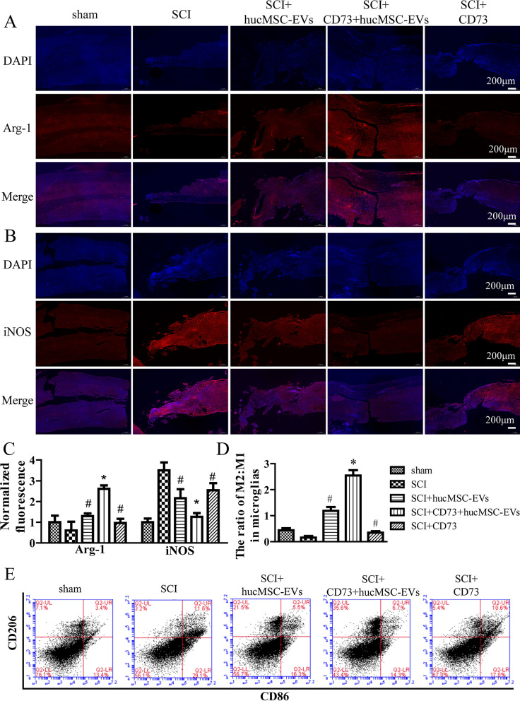
Fig. 6.
CD73+hucMSC-EVs regulate M1/M2 polarization of microglia in mice. A and B Changes of arginase-1 and iNOS are determined by immunofluorescence in different groups at × 5 magnification. C Fluorescent intensities are normalized to the sham group. (*p < 0.05 versus SCI group, #p < 0.05 versus SCI + CD73+hucMSC-EVs group, n = 5). D and E Representative dot spot of flow cytometry for microglia/macrophage subsets is shown. CD206 and CD86 are selected as biomarkers of M2 and M1 microglia, respectively. The data are calculated as M2:M1. (*p < 0.05 versus SCI group, #p < 0.05 versus SCI + CD73+hucMSC-EVs group, n = 5)

