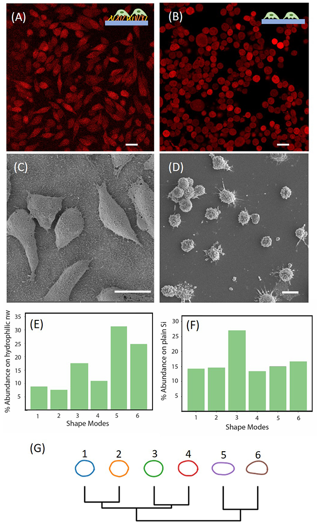
Figure 4.

(A) Fluorescence image of parental P231 cells on patterned Au/SiNWs. (B) Fluorescence image of parental P231 cells on plain Si wafer. Scale Bar 20 μm. (C) SEM image of P231 cells on the hydrophilic region of the Au/SiNWs substrate. (D) SEM image of P231 cells on flat surface. Scale bar 20 μm. (E) Distribution of various shape modes of parental P231 cells on Au/SiNWs using VAMPIRE analysis. (F) Distribution of various shape modes of parental P231 cells on plain Si wafer using VAMPIRE analysis. (G) Dendrogram of various shape modes showing the relation between each mode.
