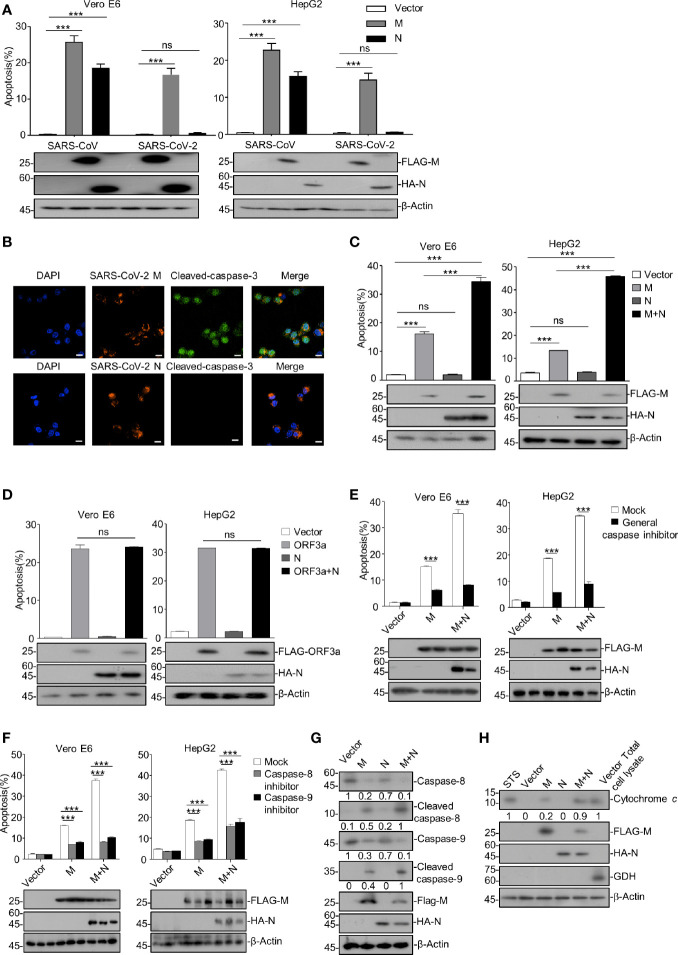
Figure 1.
SARS-CoV-2 M induces caspase-dependent apoptosis, and N specifically enhances M-induced apoptosis. (A) Vero E6 and HepG2 cells were transfected with FLAG-SARS-CoV-2 M (M), HA-SARS-CoV-2 N (N), and FLAG-SARS-CoV-2 M plus HA-SARS-CoV-2 N (M+N), also with the counterparts of SARS-CoV. After 24 h, cells were stained with Annexin V-FITC/PI for flow cytometry analysis, and the percentage of apoptotic cells was measured. (B) Vero E6 cells were transfected with FLAG-SARS-CoV-2 M or FLAG-SARS-CoV-2 N, respectively. After 24 h, cells were processed for immunofluorescence and co-stained with anti-FLAG and anti-cleaved-caspase-3 antibodies indicated. Scale bar, 20 μm. (C) Vero E6 and HepG2 cells were transfected with FLAG-SARS-CoV-2 M (M), HA-SARS-CoV-2 N (N), and FLAG-SARS-CoV-2 M plus HA-SARS-CoV-2 N (M+N). After 24 h, cells were collected for Western blotting (with anti-FLAG, anti-HA, or anti-β-Actin) or stained with Annexin V-FITC/PI for flow cytometry analysis, and the percentage of apoptotic cells were measured. (D) Vero E6 and HepG2 cells were transfected with FLAG-SARS-CoV-2 ORF3a (ORF3a), HA-SARS-CoV-2 N (N), and FLAG-SARS-CoV-2 ORF3a plus HA-SARS-CoV-2 N (ORF3a+N). After 24 h, cells were collected for Western blotting (with anti-FLAG, anti-HA, and anti-β-Actin) or stained with Annexin V-FITC/PI for flow cytometry analysis, and the percentage of apoptotic cells were measured. (E, F) Vero E6 and HepG2 cells were transfected with FLAG-SARS-CoV-2 M and FLAG-SARS-CoV-2 M plus HA-SARS-CoV-2 N and treated with DMSO, a general caspase inhibitor, a caspase-8 inhibitor, and caspase-9 inhibitor, respectively. After 24 h, cells were collected for Western blotting (with anti-FLAG, anti-HA, or anti-β-Actin) or stained with Annexin V-FITC/PI for flow cytometry analysis, and the percentage of apoptotic cells were measured. (G, H) HEK293T cells were transfected with FLAG-SARS-CoV-2 M, HA-SARS-CoV-2 N, and FLAG-SARS-CoV-2 M plus HA-SARS-CoV-2 N (STS, staurosporine as a positive control). After 24 h, cells were collected for Western blotting with antibodies to the indicated proteins. To examine the levels of cytochrome c in the cytosol, mitochondria were separated via gradient centrifugation, and cell lysate without mitochondria was subjected for Western blotting with antibodies to the indicated proteins. The total cell lysate within intact mitochondria was used as the positive control. GDH, glutamate dehydrogenase. ***P < 0.001 by two-tailed Student’s t-test. ns, non significant. The densities of blots were analyzed with ImageJ software.

