Figure 5.

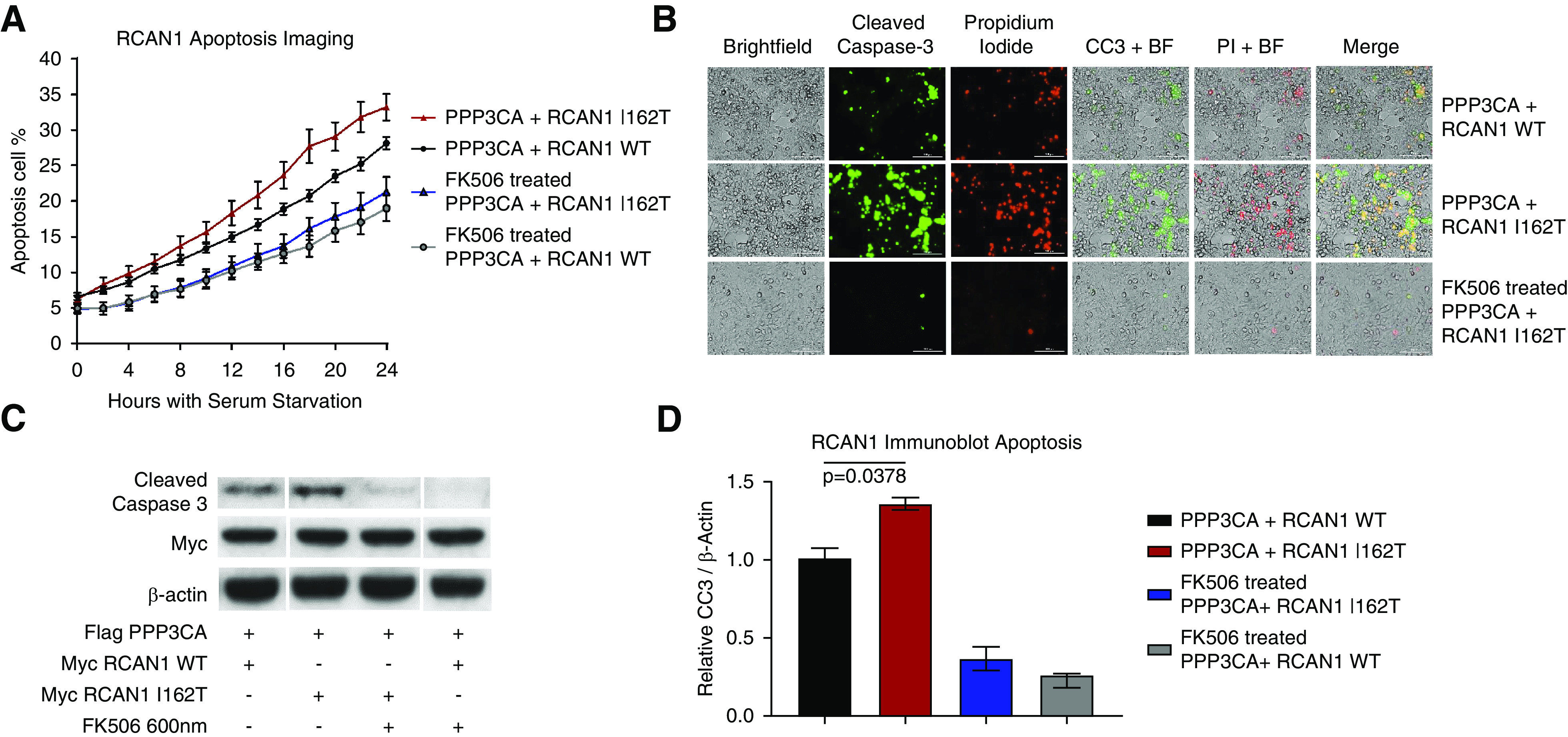
Mutant RCAN1 causes increased apoptosis that can be rescue by CNI FK506. (A) HEK293 cells were transfected with constructs containing PPP3CA (CN) and either WT RCAN1 or the p.I162T variant, and the cells were exposed to serum deprivation. We analyzed the susceptibility to apoptosis using a fluorescent reporter of caspase-3 activity over 24 hours. RCAN1 I162T–expressing cells (red) displayed increased apoptosis compared with WT RCAN1 cells (black) (P<0.02 for all time points between 18 and 24 hours, two-way ANOVA). This increased apoptosis in the RCAN1 mutants was rescued by treatment with 1 µM FK506 (P>0.3 for all time points), demonstrating this aberrant apoptosis in mutant cells is due to the increased CN activity (n>16 for all samples). (B) This increased apoptosis could be seen in still images taken 24 hours after serum starvation, which showed increased apoptosis (green, cleaved caspase-3 [CC3]) and necrosis (red, propidium iodide [PI]) in RCAN1 I162T–expressing cells compared with WT. (C and D) The increased apoptosis and rescue was confirmed through Western blot analysis of cleaved caspase-3 expression after 48 hours of serum starvation (P=0.02, n=3, one-way ANOVA). BF, bright-field imaging.
