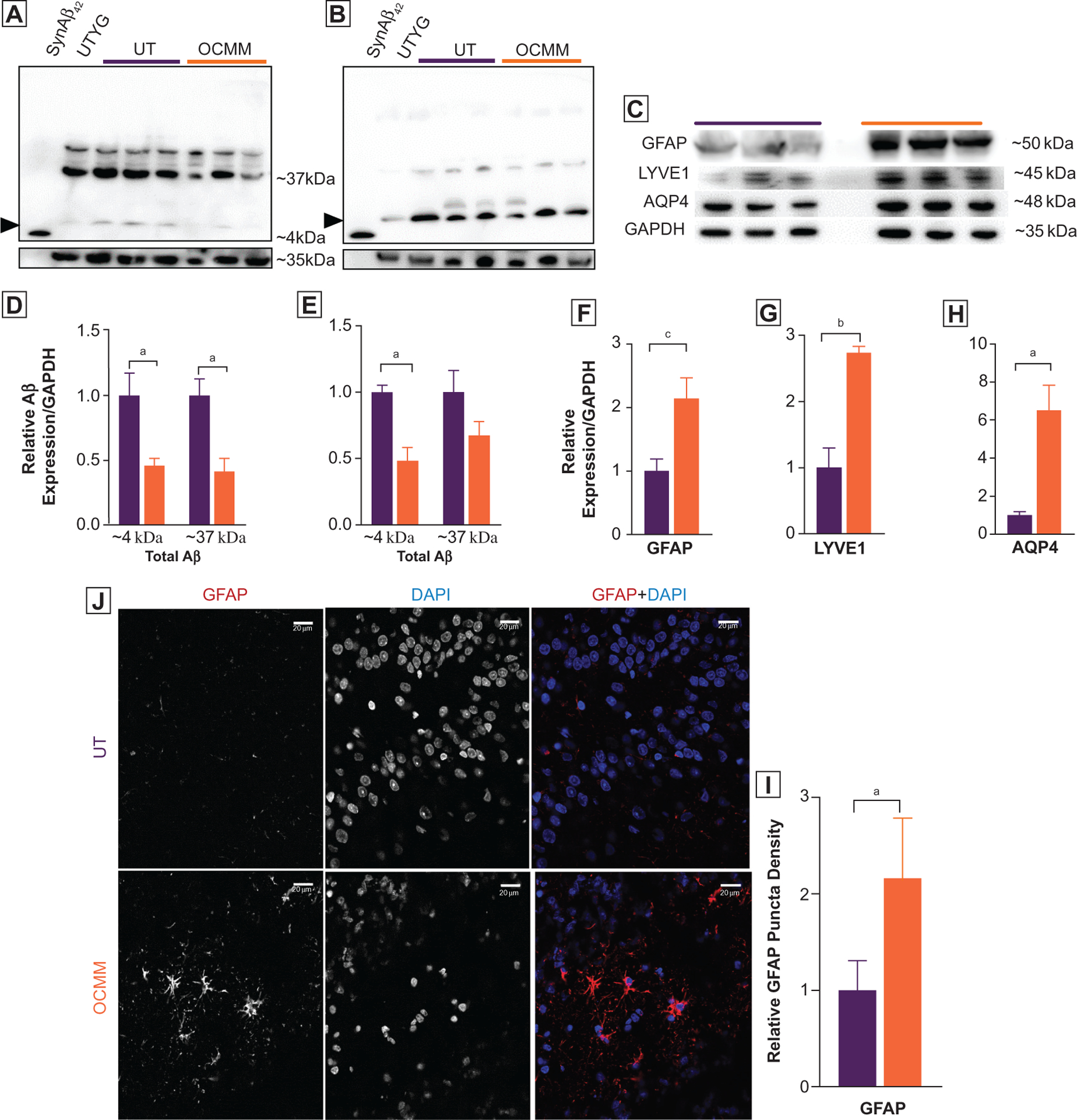
Figure 3.

(A, B) Representative whole-gel pictures of Western blot assays show the total amyloid β (Aβ) motif containing proteins as probed with 2 monoclonal anti-Aβ42 primary antibodies recognizing N-terminal (Abcam, No. ab201060 [A]) and C-terminal (Cell Signaling, No. 14974 [B]) end of the Aβ42 peptide. SynAβ42, control synthetic rat Aβ42 peptide (Anaspec No. AS-25381). Untreated young adult (UTYa), control rat brain lysate. Arrowheads indicate the expected ~4-kDa size Aβ42 peptide. Higher-molecular-weight (~37 kDa) bands indicate the presence of variable length of amyloid C-terminal domain fragments with intact Aβ42 motif. Relative density analysis revealed that rats treated with osteopathic cranial manipulative medicine (OCMM; n=3) had significantly less (P<.05, 100 μg of total protein was loaded) anticipated ~4-kDa Aβ42 peptide and ~37 kDa fragments in N-terminal ([D] P<.05 for ~4 kDa, P<.05 for ~37 kDa) and C-terminal ([E] P<.05 for ~4 kDa) recognizing anti-Aβ42 antibodies. (C) Western blots showing total glial fibrillary acidic protein (GFAP), aquaporin-4 (AQP4), and lymphatic vessel endothelial hyaluronic receptor 1 (LYVE1) expression in aged untreated (UT) and OCMM-treated rats; GAPDH served as a control. All Western blots were repeated with duplicate samples of each animal brain lysate. (F-H) Histograms of relative density analysis reveal that OCMM-treated rats had significantly higher GFAP (P<.001), AQP4 (P<.05), and LYVE1 (P<.01) expression than the UT rats (P<.05). (J) Immunohistochemical assay shows the increased expression of GFAP at the dentate gyrus in OCMM-treated rats. (I) Histograms represent the normalized averages and statistical significance. Unpaired 2-tailed t test, aP<.05, bP<.01, cP<.001. Scale bar, 20 μM.
