
Fig. 5. Formation of a localized tumor/T cell αvβ8/L–TGF-β signaling complex.
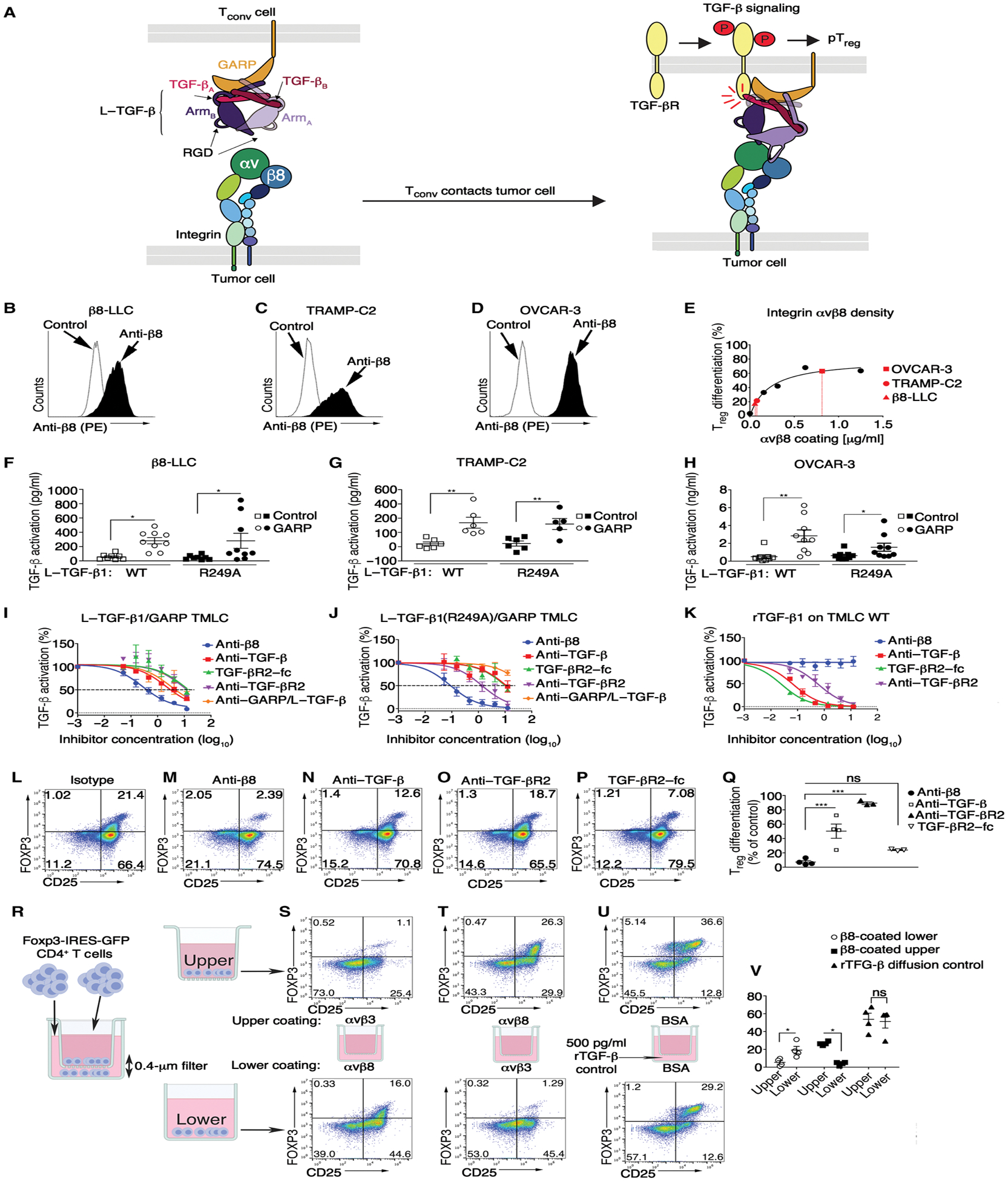
(A) Cartoon of structure-based model of αvβ8-mediated TGF-β activation and signaling based on structures of αvβ8/L–TGF-β (29, 43), L–TGF-β/GARP (51), and TGF-βR2/TGF-β1 (66). Integrin αv and β8 subunits, latency associated peptide (LAP) of dimeric L–TGF-β (subunit A + B), dimeric TGF-β (subunit A + B), TGF-βR2, and GARP color-coded matching annotations. Integrin and GARP/TGF-βR2 trans-membrane domains span tumor or Treg lipid bilayers, respectively. (B) αvβ8 surface expression in β8-LLC, (C) TRAMP-C2, and (D) OVCAR-3 stained with C6D4 (1 μg/ml) compared with iso-type. (E) Treg differentiation over a range of αvβ8 coating concentrations. Superimposed in red are β8-LLC (red triangle), TRAMP-C2 (red circle), and OVCAR-3 (red square) according to calculated αvβ8 cell surface receptor density. (F to H) WT human L–TGF-β1 or mutant incapable of producing diffusible TGF-β1 [L–TGF-β1(R249A)] expressed alone (square symbols) or coexpressed with human GARP (circles) in TGF-β reporter cells (TMLC) and cocultured with (F) β8-LLC, (G) TRAMP-C2, or (H) OVCAR-3. Outliers (Rout) were removed from (F and G). Shown is TGF-β activation (means ± SEM) determined using rTGF-β standard curve of each TMLC line (n ≥ 6). (I to K) Inhibition curves of anti-β8 (C6D4, blue line) compared with anti–pan–TGF-β (1D11, red line), TGF-βR2–Fc receptor trap (green line), anti-human GARP/L–TGF-β (MHG-8, purple line), or anti-human/mouse TGF-βR2 (clone 8322, orange line) generated using (I) WT human L–TGF-β1/human GARP TMLC, (J) human L–TGF-β1(R249A)/human GARP TMLC or control, and (K) WT TMLC cells with 500 pg of rTGF-β1. Shown is percent inhibition relative to no antibody control. Inhibitor concentrations are shown in μg/ml (log10). (L to P) iTreg differentiation of activated CD4+ T cells from foxp3-IRES-GFP splenocytes on immobilized αvβ8 in the presence of (L) isotype, (M) anti-β8 (C6D4), (N) anti–TGF-β1 (1D11), (O) anti–TGF-βR2 (clone 8322), (P) or TGF-βR2–Fc. (Q) Results enumerated in scatterplots (n ≥ 3). (R) Schematic of Transwell assay showing that diffusible TGF-β has no role in αvβ8-mediated iTreg differentiation. CD4+ T cells plated into upper and lower chambers under stimulating conditions. (S) αvβ8 coated on lower, αvβ3 control on upper, or (T) vice versa. (U) Active rTGF-β added to lower chamber media demonstrating diffusion of rTGF-β into the upper chamber inducing conversion of non-Treg CD4+ T cells (Tconv) to CD25+FOXP3+ Treg. (V) Scatterplots (n = 4) show gated CD4+ T cells stained with anti-CD25 (x axis) with FOXP3 expression determined by GFP (y axis). *P < 0.05, **P < 0.01, and ***P < 0.001 by one-way ANOVA for multiple comparisons followed by Sidak’s post-test.