Figure 6.

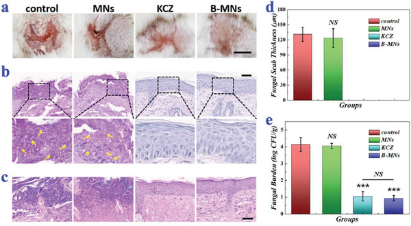
In‐vivo antifungal performances of B. subtilis‐loaded ice microneedles. a) Representative photos of back skins of the mice in the control group, the MNs group, the KCZ group, and the B‐MNs group on day 13. b) PAS staining of mouse back skins in different groups on day 13. The arrows pointed to the pseudohyphae of C. albicans. c) H&E staining of mouse back skins in different groups on day 13. d) Quantitative analysis of the fungal scab thicknesses of the mice (n = 4 for each group; Student's t‐test was conducted for comparison; NS: nonsignificant). e) Quantitative analysis of fungal burden of the infected skin tissues (n = 6 for each group; Student's t‐test was conducted for comparison; *** p < 0.001, NS: nonsignificant). Scale bars: 0.5 cm in (a) and 50 µm in (b,c).
