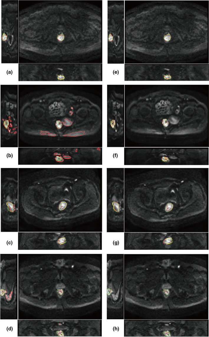
FIGURE 4.

Example of segmentation. (a–b) semi‐automatic segmentation; (e–h) deep learning segmentation. The two images in each row are from the same patient. Green color shows the contour of ground truth delineated by radiologists. Red color shows the contour of segmentation. Yellow color is the overlap of green and red contours
