. 2021 Aug 23;10:e66738. doi: 10.7554/eLife.66738

Figure 5. WM repopulating myeloid cells derive from an existing Cx3cr1+ cell source originating from the SVZ/WM area.

(A–H) Two-month-old WT mice were treated with PLX3397 (600 ppm) for 14 days, then allowed to recover without PLX3397 for 3, 5, and 7 days. (A–B) Representative 63x immunofluorescence images of proliferating (Ki67+, blue) myeloid cells (IBA1+, green) staining for positive for common cell lineage/precursor cell markers: NESTIN (red, A) and MASH1 (red, B) in the SVZ of control, 14 day PLX3397, 3 day recovery, 5 day recovery, and 7 day recovery mice. (C–E) CreER-directed lineage-specific labeling. In these mouse lines, tamoxifen-inducible Cre-recombinase is expressed under control of the promoter of interest. When activated by tamoxifen, the CreER fusion protein translocates to the nucleus allowing transient recombination to occur and, when crossed to a YFP reporter, visualization of induced expression via eYFP. (C–D) Representative 20x images of IBA1+ (red) and associated promoter-driven lineage-derived (YFP, green) cells in control and 7 day recovery Ascl1CreERT2/YFP (B) and NestinCreERT2/YFP (C) mice. (E) Representative 20x images of IBA1+ (red) and Cx3cr1+ lineage derived (YFP, green) cells in control and 7 day recovery mice. (F–H) Representative coronal (F) and sagittal (G–H) brain images of IBA1+ (green) and Ki67+ (red, B–C) cells near SVZ/RMS regions between 5 and 14 days recovery. Inserts provide higher resolution images of cells near the SVZ/RMS proliferative site of repopulation, illustrating the spread of repopulating cells via WM/axonal tracts (i.e. RMS) between the CP and OB and between other WM regions (IC and CeP). CP, caudoputamen; CC, corpus callosum; CTX, cortex; LV, lateral ventricle; RMS, rostral migratory stream; SVZ, subventricular zone; OB, olfactory bulb; IC, internal capsule; CeP, cerebral peduncle.

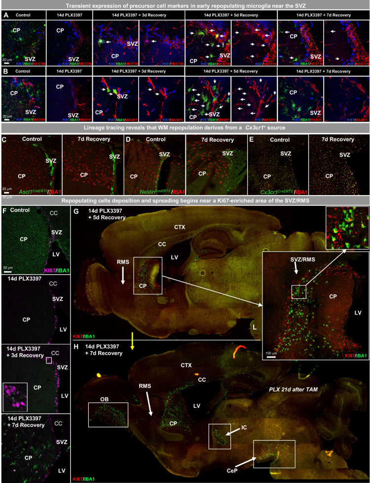
Figure 5—source data 1. WM repopulating myeloid cells derive from an existing Cx3cr1+ cell source originating from the SVZ/WM area.

Figure 5.

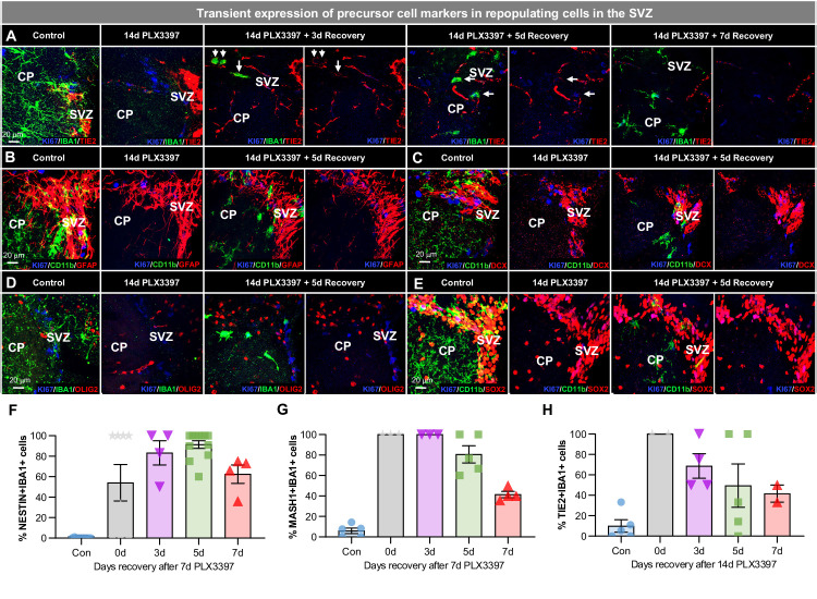
Figure 5—figure supplement 1. Early WM repopulating myeloid cells transiently express some precursor cell markers.

Figure 5—figure supplement 1.

(A–E) Representative 63x immunofluorescence images of proliferating (Ki67+, blue) myeloid cells (CD11b+ or IBA1+, green) staining for positive for common cell lineage/precursor cell markers: TIE2 (red, A), and negative for: GFAP (red, B), doublecortin (DCX, red, C), OLIG2 (red, D), and SOX2 (red, E) in the SVZ of control, 14 day PLX3397, 3 day recovery, 5 day recovery, and 7 day recovery mice. (F–H) Quantification of IBA1+ cells that co-stain with the precursor cell markers: NESTIN (F), MASH1 (G), and TIE2 (H). Data are represented as mean ± SEM (n=4–5). CP, caudoputamen; SVZ, subventricular zone.
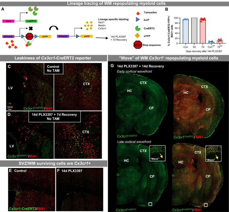
Figure 5—figure supplement 2. WM repopulating myeloid cells derive from an existing Cx3cr1+ cell source originating from the SVZ/WM area.

Figure 5—figure supplement 2.

(A–G) CreER-directed lineage-specific labeling. (A) In these mouse lines, tamoxifen-inducible Cre-recombinase is expressed under control of the promoter of interest. When activated by tamoxifen, the CreER fusion protein translocates to the nucleus allowing transient recombination to occur and, when crossed to a YFP reporter, visualization of induced expression via eYFP. In this study, mice received 14 days of PLX3397 treatment 21 days following last tamoxifen injection. (B) Quantification of Cx3cr1CreERT2-YFP+IBA1+ cells in control, 14 day PLX3397, and 7 day recovery mice, in the presence and absence of tamoxifen treatment. (C–D) Representative images of Cx3cr1CreERT2-YFP+ (green) and IBA1+ (red) cells in control (C) and 7 day recovery (D) mice treated without tamoxifen. (E) Representative tile scan confocal images of Cx3cr1CreERT2-YFP+ (green) and IBA1+ (red) cells in control and 14 day PLX3397 mice. (I) Representative whole brain images of Cx3cr1CreERT2-YFP+ (green) and IBA1+ (red) cells in 14 day recovery mice, highlighting the ‘wave.’ CTX, cortex; CP, caudoputamen; HC, hippocampus; LV, lateral ventricle; SVZ, subventricular zone.