. 2021 Jun 29;10:e57245. doi: 10.7554/eLife.57245

Figure 4. Lead effects pn mouse primary photoreceptor cells - thapsigargin-induced cell death.

(A) Primary photoreceptor survival assay protocol. (B) Box plots of photoreceptor survival effects of lead compounds and the positive control compound (POS). Statistically significant survival effects are marked with an asterisk. Survival effects, adjusted p-values, and sample sizes (N) for each condition are shown in the table below. Adjusted p-values were calculated by performing Mann-Whitney U tests followed by false discovery rate (FDR) correction for multiple comparisons. Each assay consisted of six biological replicates per condition and a minimum of two experimental repeats was performed across all conditions (Figure 4—source data 1). (C) Representative images from each condition. Lead compound abbreviations: as in prior figures. Others: EthD, Ethidium homodimer; GFP, green fluorescent protein.

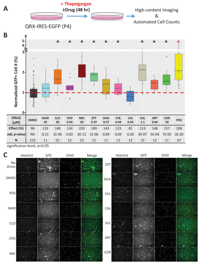
Figure 4—source data 1. Compound tests of mouse photoreceptor culture - thapsigargin assay.

Figure 4.

Figure 4—figure supplement 1. Lead effects on mouse photoreceptor cultures - tunicamycin-induced cell death.

Figure 4—figure supplement 1.

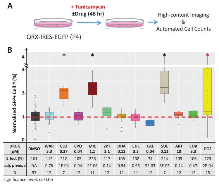
A) Diagram of primary photoreceptor survival assay protocol. Primary retinal cells were isolated from QRX mice and cultured for 2 days in tunicamycin (an ER stressor that induces cell death) and lead compounds or DMSO (control). After 48 hr, cells were stained with Hoescht and ethidium homodimer followed by high-content imaging and automated cell counting to assess the survival effects of lead compounds on GFP-expressing photoreceptor cells. Cell counts for each condition were normalized to the DMSO controls on the same plate. (B) Box plots of photoreceptor survival effects of lead compounds and the positive control compound. Conditions resulting in statistically significant survival effects are marked with an asterisk. The table below shows survival effects (normalized % GFP-expressing cells), adjusted p values, and total sample sizes (N) for each condition. Each assay consisted of six biological replicates per condition and a minimum of two experimental repeats was performed across all conditions (Figure 4—figure supplement 1—source data 1). Adjusted p values were calculated by performing Mann-Whitney U tests followed by false discovery rate (FDR) correction for multiple comparisons. Lead compound abbreviations: WAR, Warfarin; CLO, Cloxyquin; CPO, Ciclopirox olamine; MIC, Miconazole; ZPT, Zinc pyrithione; DHA, Dihydroartemisinin; CHL, Chloroxine; CAL, Calcimycin; SUL, Sulindac; ART, Artemisinin; COR, Cortexolone. Other abbreviations: CI, confidence interval; DMSO, dimethylsulfoxide; POS (positive control).
Figure 4—figure supplement 1—source data 1. Quantification of mouse photoreceptor assay - tunicamycin-induced cell death.
Figure 4—figure supplement 2. Paired lead compound survival effects in mouse photoreceptor cultures.

Figure 4—figure supplement 2.

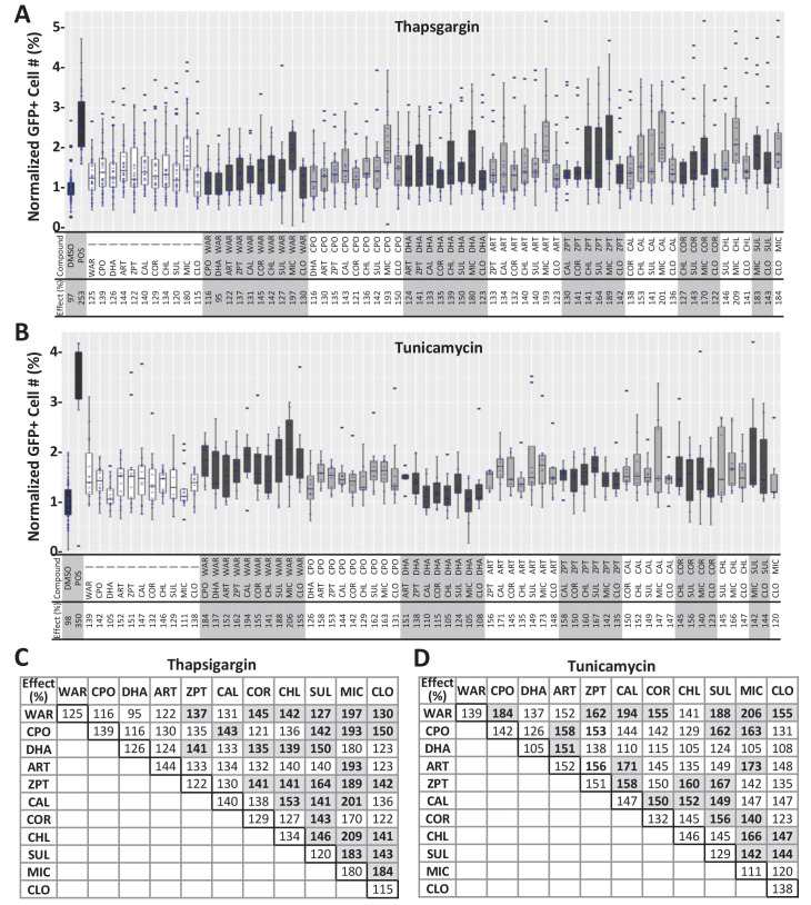
(A–B) Lead compounds were tested individually and in pairs for survival effects in primary photoreceptor cells isolated from QRX mice as per Figure 4. Box plots and summary tables (below plots) show relative survival effects in thapsigargin-treated (A) and tunicamycin-treated (B) cultures across all conditions. (C–D) Tables summarize survival effects of individual compounds (outlined boxes on diagonal) and paired compounds for thapsigargin-treated (C) and tunicamycin-treated (D) cultures, trend toward enhanced survival effects are indicated by bolded numbers, gray boxes (median effects). Mann-Whitney U tests followed by Bonferroni correction for multiple comparison (α=0.0006 adjusted significance level) showed that enhanced effects of paired conditions were not statistically significant relative to individual compound controls. (Figure 4—figure supplement 2—source data 1). Lead compound abbreviations: WAR, Warfarin; CLO, Cloxyquin; CPO, Ciclopirox olamine; MIC, Miconazole; ZPT, Zinc pyrithione; DHA, Dihydroartemisinin; CHL, Chloroxine; CAL, Calcimycin; SUL, Sulindac; ART, Artemisinin; COR, Cortexolone. Other abbreviations: DMSO, dimethylsulfoxide; POS (positive control).
Figure 4—figure supplement 2—source data 1. Paired drug tests - mouse photoreceptor cultures.