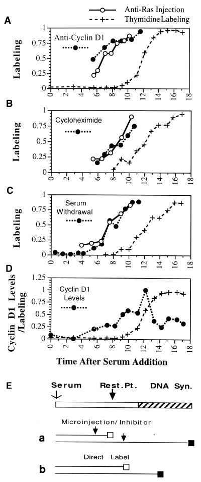
FIG. 1.
Restriction point determination in quiescent cells. Inhibitory treatments were applied at the indicated times following serum addition to quiescent NIH 3T3 cells. Thymidine was added following antibody microinjection (or addition of the inhibitor) until 24 h following the initial addition of serum to the culture, at which time the cells were fixed and autoradiographed. The proportion of cells which were labeled with thymidine following treatment is plotted versus the time (in hours) following serum addition at which the injection (inhibitory treatment) took place. To identify the time these cells first enter S phase, labeled thymidine was added together with serum to a parallel set of cultures, which were fixed at the indicated times. (A to C) In each experiment, an inhibitor as indicated (solid circles in each panel) was analyzed together with anti-Ras injection (○), while thymidine labeling (+) was performed in each experiment to determine the timing of entry into S phase. (D) Accumulation of nuclear cyclin D1 protein determined by antibody staining at the indicated times following serum addition to quiescent cells, reported as the proportion of maximal antibody staining at each time point. For each point, the average of approximately 100 individual cells is given. (E) Procedures used. The top bar represents cell cycle progression following serum addition from G0 phase into S phase; the restriction point (Rest. Pt.) is indicated together with DNA synthesis. The lower lines indicate progression of cells through the cell cycle. An open box indicates the cell cycle point to which the cell had progressed, and a solid box indicates thymidine labeling. (a) Treatment with inhibitors or injection of antibody. Cells treated prior to the restriction point (arrow, top line) would be unable to progress past the restriction point and thus unable to incorporate thymidine. Treatment after the cells had passed the restriction point (arrow, lower line) would be unable to inhibit entry into S phase and would result in thymidine labeling of the cells. Direct labeling of cells at the time of serum addition (b) would result in thymidine labeling of any cell in S phase at the time the cells were fixed.

