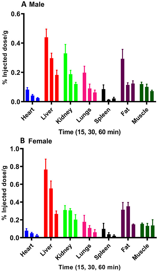
Figure 3.
Biodistribution of [3H]MCL-536 in peripheral organs in male and female rats. [3H]MCL-536 (6 μCi/300 g body weight) was injected into the tail vein; 15, 30, and 60 min after injection, peripheral organs were dissected, and the tissue weighed and dissolved in 1 mL of 0.8 N NaOH. After addition of 5 mL of scintillant, samples were counted in a β-counter and the %ID/g wet tissue was calculated. Data are presented as mean ± SEM of 4 animals per time point and ligand. (A) Male rats. (B) Female rats.

