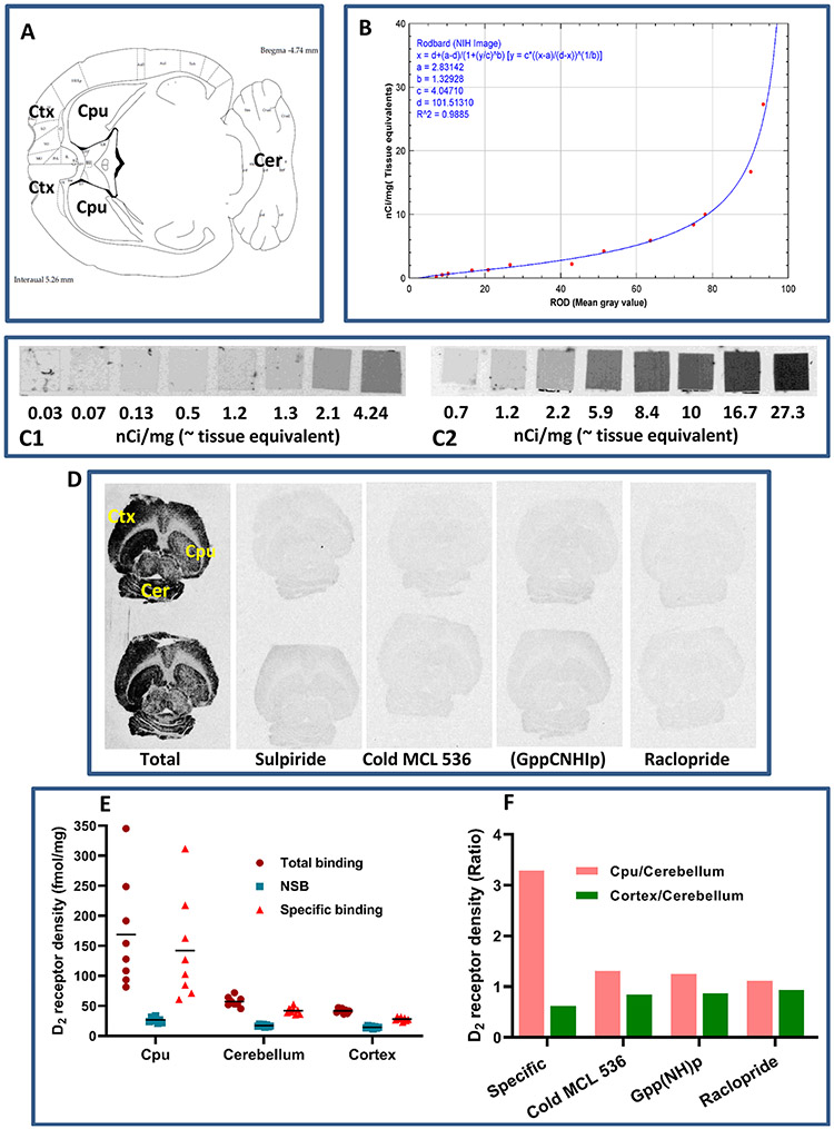
Figure 4.
In vitro autoradiography showing the binding of [3H]MCL-536 in the rat brain. Transaxial brain sections were preincubated with buffer to eliminate nonspecific binding. Afterward, sections were incubated with [3H]MCL-536 alone or in combination with D2 receptor antagonists or cold MCL-536 to show specificity. Dried sections were exposed to autoradiography film for visualization. (A) Schematic of a transaxial brain section; CPU, caudate putamen; Ctx, cortex; Cer, cerebellum. (B) Conversion of gray scale values to relative optical densities (ROD). (C) 3H microscales were coexposed on the same films as the experimental samples to allow quantification. (D) In vitro binding of radioligand to 10 μm thick rat brain transaxial slices in the presence or absence of 10 M sulpiride, 100 nM cold ligand, 200 μM Gpp(NH)p, or 100 nM raclopride. (E) Quantification of receptor density for D2 (fmol/mg). (F) D2 receptor density ratio in striatum versus cerebellum and cortex versus cerebellum in the presence or absence of other radioligands or cold MCL 536.

