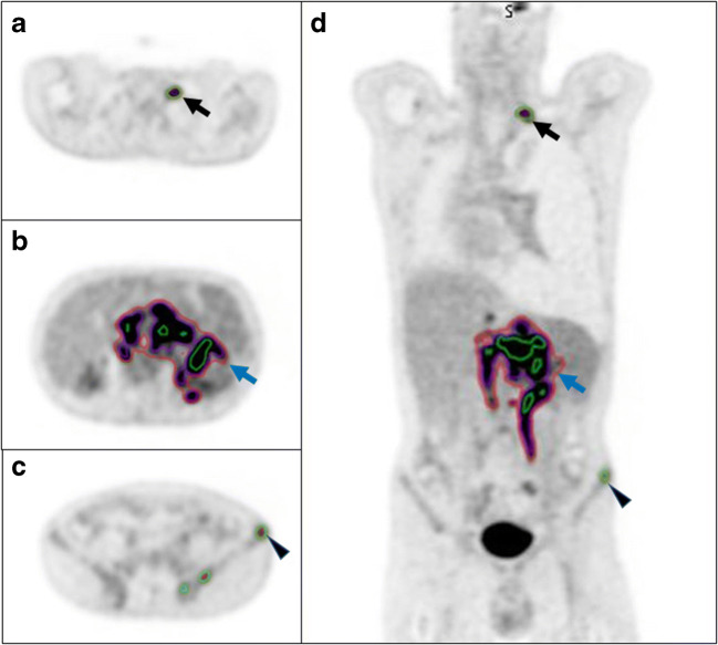
Fig. 8.
Select axial (a–c) and coronal slices (d) from an FDG PET/CT study from a patient with DLBCL demonstrating three different contouring methods (green = 41% SUVmax; red = 1.5 x SUVmean of the liver; purple = 4.0 SUV). For smaller lesions, the 41% SUVmax contour is larger than the other two methods, black arrow and arrowhead. For larger more heterogenous lesions, the 41% SUVmax is the smallest of the three contours (blue arrow)

