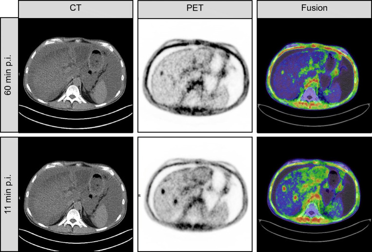
Fig. 3.
Case example of a patient with discrepant findings between early imaging and late imaging. This is a case example of a 54-year-old patient with metastatic pancreatic cancer. FAPI-46 PET was performed for restaging after resection and de novo peritoneal involvement. FAPI-46 PET performed 11 min after injection revealed two lesions of which one was not noted on late imaging. Increased background uptake is noted due involvement of hepatic viscera with possible intrahepatic cholangitis. Diffuse peritoneal involvement was noted on both scans

