Figure 1.
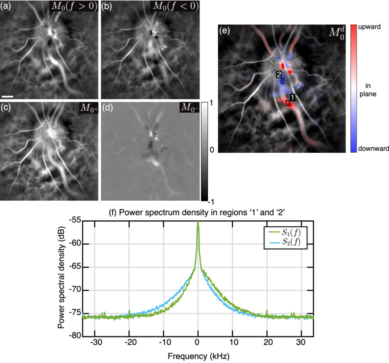
Directional blood flow contrast. (a,b) Power Doppler images from the positive and negative parts of the Doppler spectrum. (c,d) Sum and difference of those two images. (e) Directional power Doppler image on which upward and downward flow give red and blue contrasts, respectively. (f) Doppler power spectrum density in two regions of a blood vessel where the local flow has upward (green trace) and downward components (blue trace), respectively.

