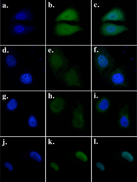
Fig. 1. Cellular localization of GFP fusions in HeLa cells transiently transfected with a vector expressing GFP (a, b, c), PB-GFP (d, e, f), PB.1–558-GFP (g, h, i) and PB.NLS-1–558-GFP (j, k, l).
The left panels (a, d, g, j) show the nuclear genomic DNA staining by Hoechst 33342, the middle panels (b, e, h, k)) show GFP fluorescence, the right panels (c, f, I, l) correspond to merge pictures.

