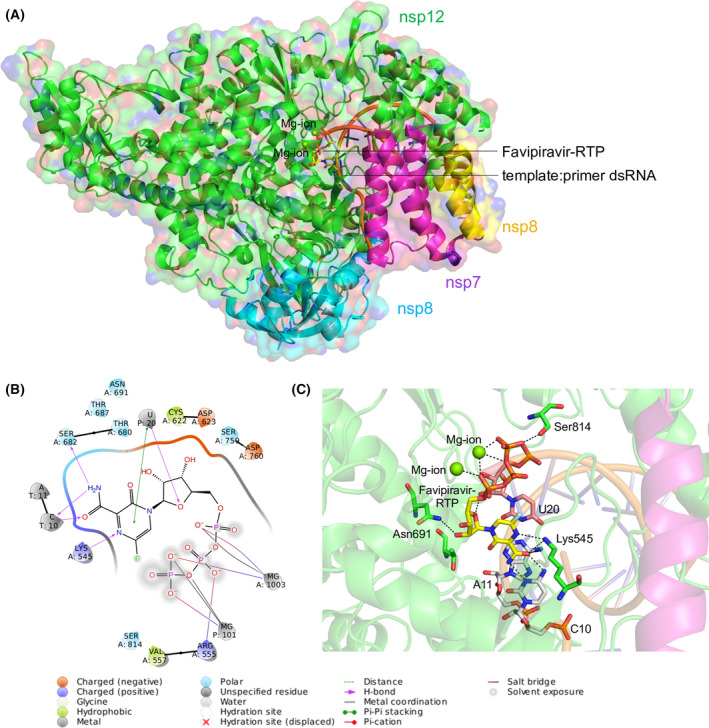
Fig. 1.

Structure of the SARS‐CoV‐2 RdRp complex. (A) The cryo‐EM structure of SARS‐CoV‐2 RdRp in complex with the template:primer dsRNA and favipiravir‐RTP. The nsp7, nsp8 and nsp12 domains are labelled with respective colours, and catalytic Mg2+ ion is shown. The template:primer dsRNA and favipiravir‐RTP are shown as a cartoon and stick model respectively. (B) Ligand interaction diagram of favipiravir‐RTP bound to the template:primer dsRNA. (C) Intermolecular interactions and interacting residues of nsp12 and RNA bases of favipiravir‐RTP (yellow) and Mg2+ (green sphere) coordinating the favipiravir‐RTP. Hydrogen bonds are shown as black dashes.
