
FIGURE 1.
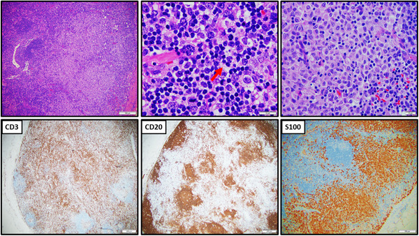
Biopsy of left axillary lymph node shows marked follicular and interfollicular hyperplasia with sinus histiocytosis and focal dendritic cells and Langerhans cells proliferation (S100+). By immunocytochemistry, there are normal T‐cells (CD3+) and B‐cells (CD20+) compartmentalization. Occasional hemophagocytosis is noted (arrow)