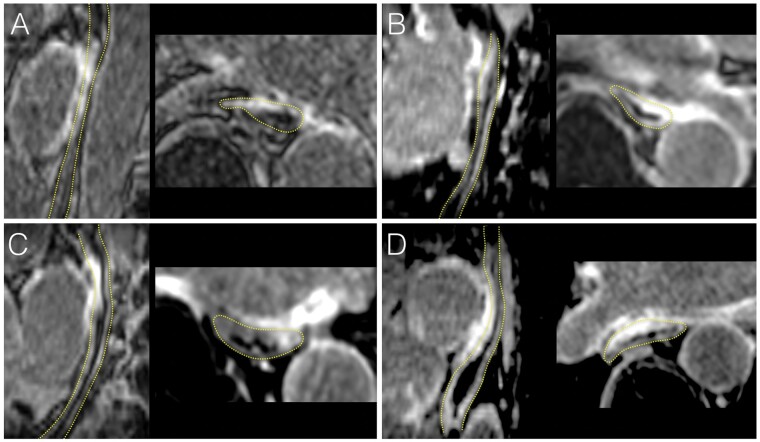
Figure 1.
Examples of acute oesophageal injuries on CMR following thermal ablation. LGE CMR images acquired less than 3 h post-ablation are shown in four patients treated with RF (A, C) or cryoballoon (B, D). In each, the oesophagus is shown in a sagittal oblique view parallel to the oesophagus (left image), and in a transaxial view (right image). The dotted yellow lines indicate oesophagus location. All patients show intense and transmural oesophageal LGE in direct contact with atrial areas targeted by ablation, also exhibiting LGE. CMR, cardiac magnetic resonance; LGE, late gadolinium enhancement; RF, radiofrequency.

