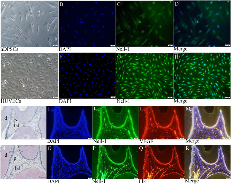
FIGURE 5.
Nell-1 distribution in DPSCs (A–D), HUVECs (E–H), and normal pulp tissues (I–R). (A,E) Normal cell feature of DPSCs and HUVECs. (B,F) DAPI: nucleus stain. (C,G) green fluorescence can be observed in the nucleus of DPSCs and HUVECs. (D,H) Merged images. (I,N) HE staining displayed the morphology and structure of normal rat pulp tissues. (K,L,P,Q) Nell-1, VEGF and Flk-1 can be found in odontoblasts, pulp fibroblasts, and endothelial cells of the blood vessels. (J,O) DAPI: nucleus stain. (M,R) Merging Nell-1 with angiogenetic markers. d, dentin; p, dental pulp; bd, blood vessel.

