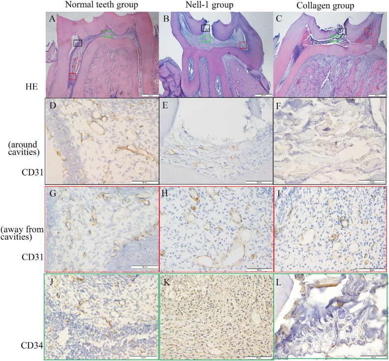
FIGURE 6.
Histology and immunohistochemistry staining. (A) Normal teeth group. (B) Nell-1 group. (C) Collagen group. HE staining showed the cavities, inflamed tissue, and normal structure of rat pulp tissues. The staining of CD31 (D–I) and CD34 (J–L) revealed the blood vessels in pulp tissues. Nell-1 induction group had higher number of blood vessels around cavities than the collagen group, and both of them had fewer blood vessels than negative control group (D–F,J–L). There was no significant difference in the area away from the cavities between three groups (G–I). d, dentin; p, dental pulp; bd, blood vessel.

