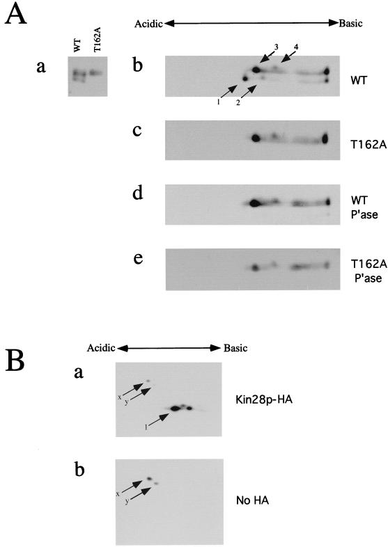
FIG. 2.
2-D gel analysis of Kin28p. (A) Protein samples containing Kin28p alleles were subjected to isoelectric focusing with a 1:1 mixture of pH 4 to 8 and pH 5 to 7 carrier ampholytes, run on SDS–12.5% polyacrylamide gels in the second dimension, and immunoblotted. To aid in identifying spots, lanes containing Kin28p and Kin28pT162A samples were run alongside tube gels in the second dimension (a). Kin28p alleles tested included wild-type (WT) Kin28p (b), Kin28pT162A (c), phosphatase (P’ase)-treated wild-type Kin28p (d), and phosphatase-treated Kin28pT162A (e). (B) To rule out the possibility of a cross-reacting species in these immunoblots, extracts from cells expressing tagged (a) or untagged (b) Kin28p were subjected to isoelectric focusing with pH 4 to 8 carrier ampholytes and processed as described for panel A.

