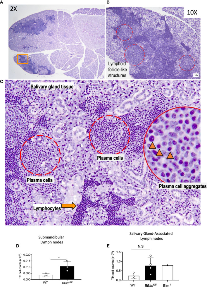
Figure 4.
Salivary glands in BBimfl/fl mice display ongoing immune reaction. (A) Salivary gland from an 8.5 month old BBimfl/fl mouse showing aggregates of lymphoid infiltrates (H&E, 2x magnification). (B) Region of salivary gland indicated by an orange rectangle in (A), showing pale areas with of lymphoid follicle-like structures (red dotted circles, 10x). (C) Higher magnification (20x) of one of these areas demonstrates plasma cell aggregates (red dotted circles); plasma cells are identified by ovoid cells with abundant pale basophilic cytoplasm and eccentric nucleus (inset, black arrowheads, 40x), adjacent to lymphocytic infiltrates identified by smaller cells with scant cytoplasm and prominent dark round nucleus (orange arrow). (D) Numbers of Tfh cells in the submandibular lymph nodes and (E) salivary gland associated lymph nodes. Not significant (N.S) P > 0.05, *P ≤ 0.05, calculated by Students T- test.

