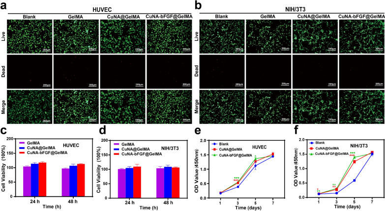
Fig. 5.
Biocompatibility evaluation of HUVEC and NIH/3T3 cells cultured in ionic extraction from 5% wt GelMA composite hydrogels (a, b) Live/dead staining of HUVEC and NIH/3T3 cells cultured in the complete medium with different ionic extraction for 24 h. Cell viability for HUVEC (c) and NIH/3T3 cells (d) was determined by CCK-8 method after cultured with different ionic extraction for 24 and 48 h. Cell proliferation assay for HUVEC (e) and NIH/3T3 cells (f) after cultured with different ionic extraction for 1, 3, 5, 7 days. Scale bar is 200 μm. Data are expressed as mean ± SD (n = 3). (*p < 0.05, **<0.01, **<0.001 compared to the blank)

