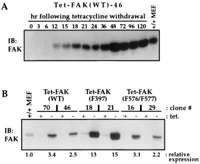
FIG. 1.
Inducible FAK expression in Tet-FAK cells. (A) Induction time course. Parallel cultures of exponentially growing Tet-FAK(WT)-46 cells were induced by tetracycline withdrawal (0 h), and cell lysates were prepared at the times indicated for analysis of FAK expression. (B) Expression levels. The indicated Tet-FAK clones were either maintained in the presence of tetracycline (+ tet.) or induced by tetracycline withdrawal for 2 days (− tet.), and then total cell lysates were prepared and analyzed for relative FAK expression levels. For both panels A and B, cells were lysed in RIPA buffer, and 30 μg of total protein was loaded per lane for assessment of FAK levels by immunoblotting (IB) with C-20 antibody and detection with 125I-labeled protein A. Control samples (MEF +/+ lanes) contained 30 μg of total protein prepared from normal mouse embryo fibroblasts. Relative expression levels were quantitated by phosphorimage volume integration.

