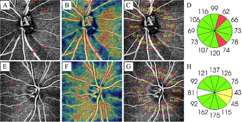
Fig. 2.
Example of PACG eye with decreased infratemporal and supratemporal vessel density of optic disc. The infratemporal and supratemporal peripapillary vessel density of glaucoma decreased and the peripapillary retinal nerve fiber layer in corresponding areas atrophied. (first row) On the contrary, these areas were very high in healthy eyes. (second row) (A) Nerve Head scan of glaucoma eye and (B) Corresponding color-coded vessel density map of the Nerve Head layer (C) Vessel density value at clock-hour region of glaucoma eye (D) Peripapillary retinal nerve fiber layer thickness at clock-hour region of glaucoma eye (E) Nerve Head scan of healthy eye and (F) Corresponding color-coded vessel density map of the Nerve Head layer (G) Vessel density value at clock-hour region of healthy eye (H) Peripapillary retinal nerve fiber layer thickness at clock-hour region of healthy eye

