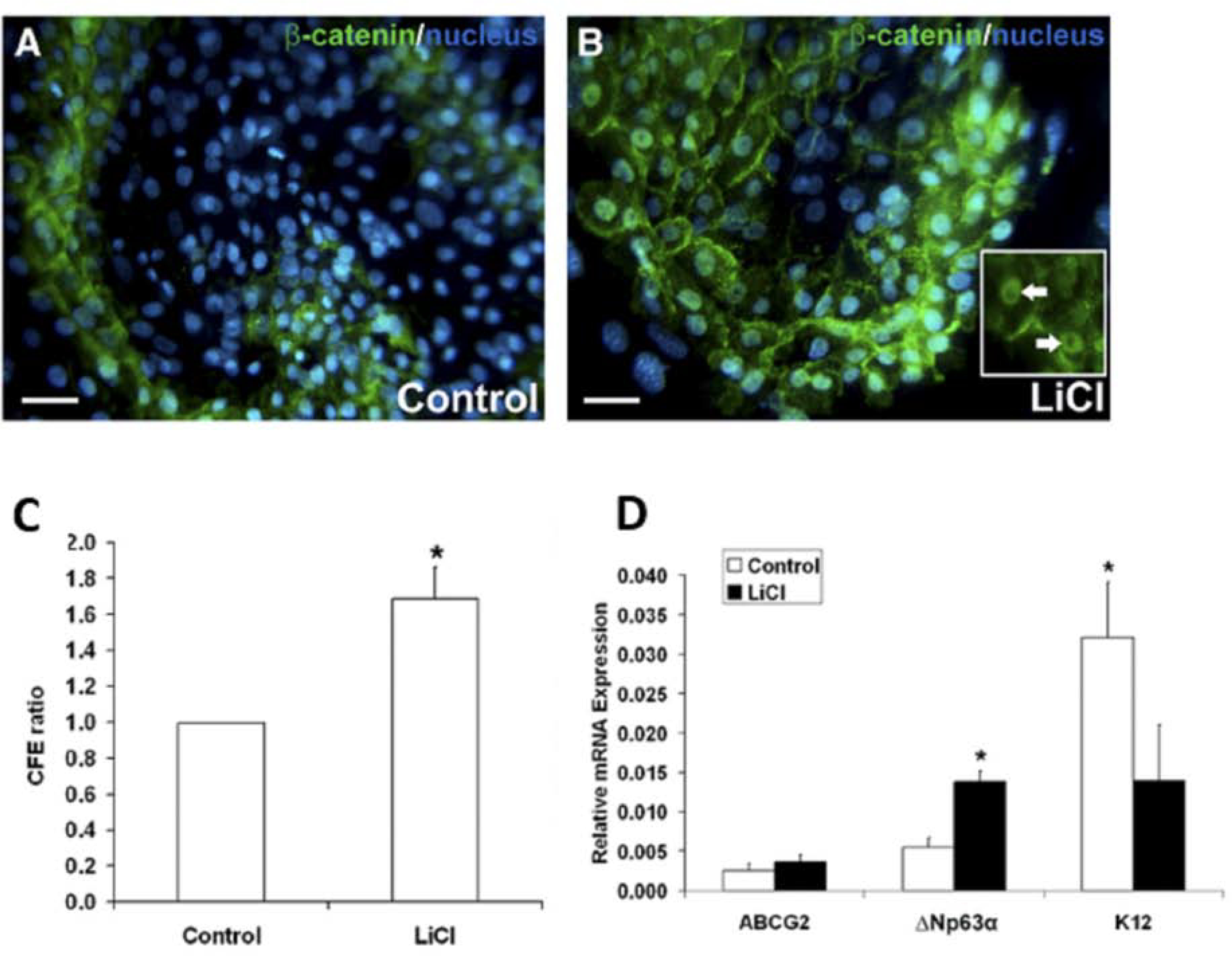
Figure 5. Activation of canonical Wnt/β-catenin signaling improves progenitor cell phenotype, while inhibition of canonical Wnt/β-catenin signaling causes loss of the stem/progenitor cell population in cultivated LSCs.

A. and B. β-catenin (green) immunofluorescent staining and Hoescht (blue) nuclear staining of LiCl-treated LSC colonies compared to control. In the LiCl-treated LSC colonies, nuclear β-catenin was observed (white arrows) that was not present in the control colonies. C. Quantification of CFE of LSC colonies as a ratio of LiCl-treated LSC cultures relative to their donor-matched control. D. Quantitative real-time PCR measure analysis of the progenitor cell markers ABCG2 and ΔNP63α, and the differentiated cell marker K12. White bars: control cultivated LSCs. Black bars: LiCl-treated cultivated LSCs. Data are represented as mean ± SEM, where *p < 0.05 was considered significant. This figure has been adapted with permission from an IOVS article (Nakatsu et al., 2011).
