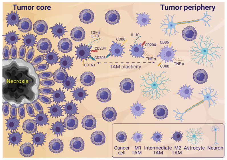
Figure 1.
Simplified glioblastoma microenvironment illustrating a section of a tumor with both tumor core and peripheral areas. In the tumor core, a necrotic area surrounded by pseudopalisading cells is shown. Necroses surrounded by pseudopalisading cells is a glioblastoma hallmark. Anti-inflammatory M2-like tumor-associated microglia and macrophages (TAMs) are mainly infiltrating the tumor core, and pro-inflammatory M1-like TAMs are mainly infiltrating the tumor periphery. Intermediate TAMs co-expressing M1 and M2 markers are also observed in the tumor microenvironment (TME). In the periphery, there is a higher presence of non-cancer cells like astrocytes and neurons, while the tumor core is more dense with cancer cells and TAMs. More cells are present in the TME—this is a simplified illustration. Abbreviations: transforming growth factor beta (TGF-β), interleukin (IL), tumor necrosis factor alpha (TNF-α). Created with BioRender.com (accessed on 16 August 2021).

