Figure 6.
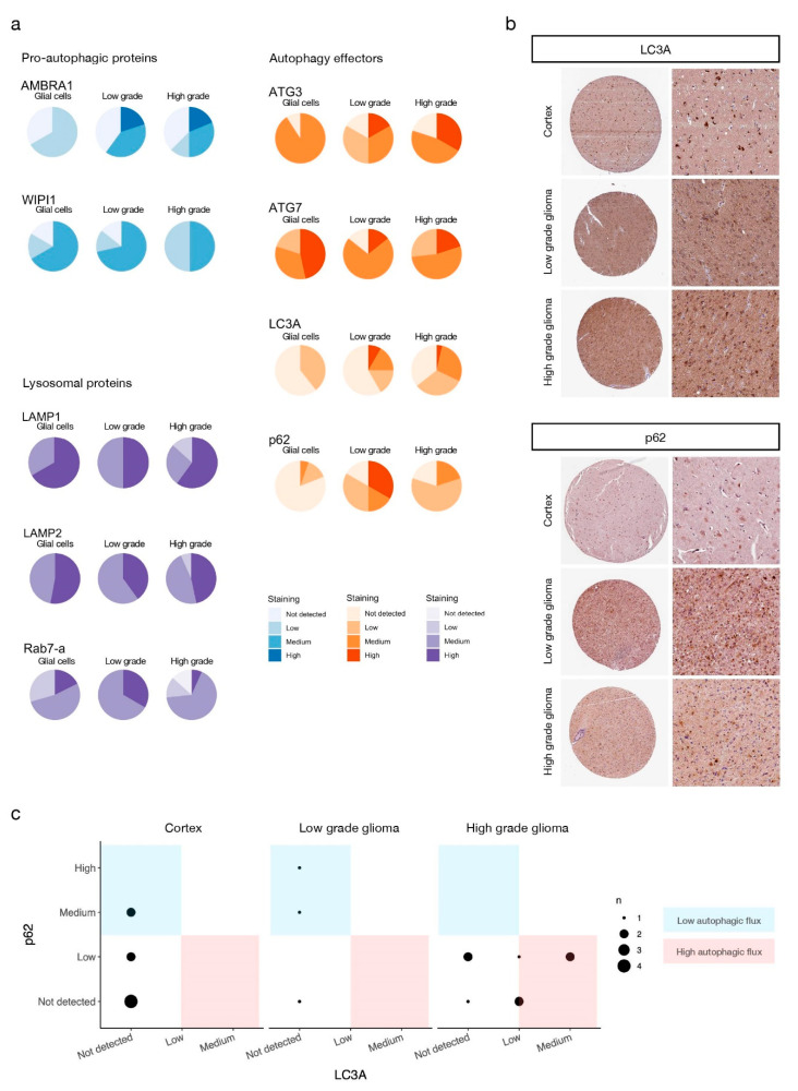
Autophagy mediators are over-expressed at the protein level in high grade glioma patients. (a) Staining levels of the indicated proteins evaluated by IHC of human healthy brain, low-grade, and high-grade glioma tissue microarrays available from the HPA (see Materials and Methods). Antibodies used for staining and patient data can be found in Tables S1–S9. IHC staining levels were manually annotated by the HPA specialists. (b) Representative IHC microarrays from the HPA (https://www.proteinatlas.org, accessed on 11 May 2021) stained for the indicated proteins. Staining levels of selected samples for LC3A: cortex–low staining (glial cells), low- and high-grade glioma–medium staining. Staining levels of selected samples for p62: cortex–not detected (glial cells), low-grade glioma–high staining, high-grade glioma–low staining. (c) Autophagic flux of IHC samples (see main text and Materials and Methods for details).

