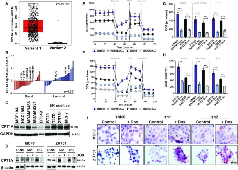
Figure 2.
CPT1A mediates FAO in ER+ breast cancer. (A) Isoform analysis of RNAseq data from 817 tumors from the TCGA demonstrated increased expression of variant 1 with >99.2% of sequenced RNA called as variant 1 on a per tumor basis (P < 0.0001). (B) Gene expression analyses of 51 breast cancer cell lines (GSE12777) demonstrates that CPT1A is more highly expressed (z-score) in ER+/luminal subtype cell lines compared to basal-like cell lines (P = 0.001). (C) Western blot analyses confirm increased Cpt1a expression in ER+ cell lines. (D) MCF7 (MCF7sh1 or MCF7sh2) or ZR751 (ZR751sh1 or ZR751sh2) cells expressing one of two independent tet-inducible shRNA show a significant decreased in Cpt1a expression by western blot following dox treatment (2 μg/ml, 96 h); no effect on MCF7shNS or ZR751shNS cells was observed upon dox treatment. (E) MCF7sh1 and (F) MCF7sh2 cells show decreased OCR levels following shRNA-mediated silencing of CPT1A under normal (DMEM; blue scale) or starvation (HBSS; gray scale) growth conditions. (G) MCF7sh1 and (H) MCF7sh2 cells show decreased basal OCR, maximum respiration and ATP (P < 0.0001) following shRNA-mediated silencing of CPT1A; the mean of four replicates with standard error are shown for each sample (Student’s t-test, ***p<0.0005). (I) MCF7 or ZR751 CPT1A shRNA expressing cells show increased lipid droplet accumulation after dox (2 μg/ml, 96 h) treatment; no effect was observed in shNS expressing cells.

