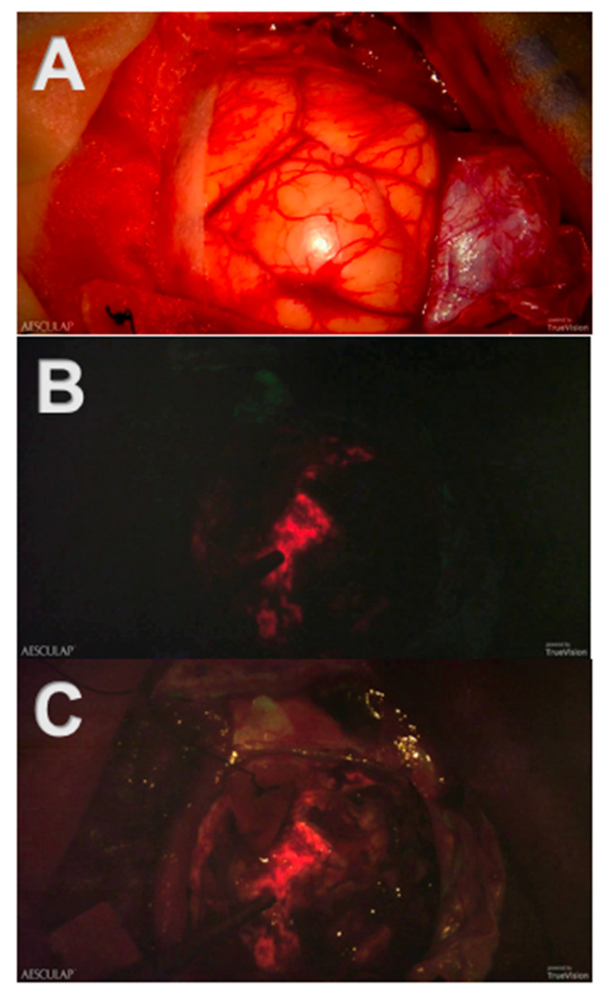
Figure 3.
Screenshots of intraoperative recordings with Aeos during resection of a glioblastoma multiforme assisted by 5-ALA-induced PpIX fluorescence: (A) Conventional white light. The tumor and its margins are elusive under conventional white light. (B) Blue light. The tumor is clearly visible in strong fluorescence under dimmed white light and activated blue light. (C) Simultaneous usage of the white and blue light source: Tumor margins are easier to identify.

