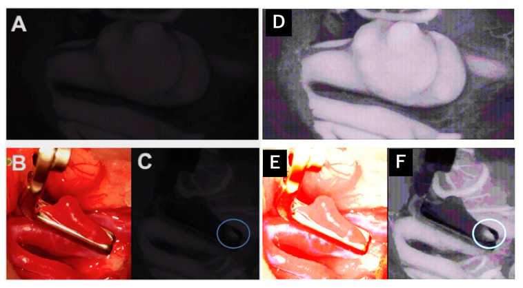
Figure 4.
Intraoperative administration of indocyanine green during aneurysm clipping: Perception of indocyanine green during Aeos surgery in A and C. The unclipped aneurysm of the medial cerebral artery is dissected and visualized in (A). Visualization of the aneurysm under conventional light during surgery closed with a right-angled Sugita clip in (B). In (C) the light of the Aeos is on for post clipping control with indocyanine green. Rest perfusion in the clipped sack of the aneurysm at the right angle is highlighted with indocyanine green. Figure (D–F) show even enhanced contrast by image processing, which however intraoperativly is not necessary due to high definition backlighted LED screen.

