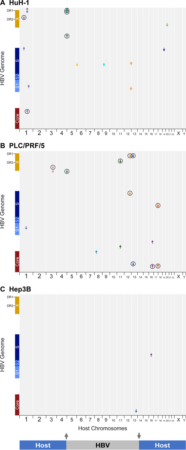
FIG 1.

Short-read sequencing defines locations and transcriptional activity of integrated HBV in HCC cell lines. (A) huH-1, (B) PLC/PRF/5, and (C) Hep3B cell lines were analyzed by short-read RNA-Seq and WGS. HBV integrations were identified by chimeric HBV-host junctions and their locations mapped as arrows according to the aligned sequences to the HBV genome (y axis) and host genome (x axis). Each arrow color corresponds to an individual human chromosome, and its direction correlates to the orientation of the HBV sequence within the associated chimeric read. Circles indicate junctions that were also detected by RNA-Seq.
