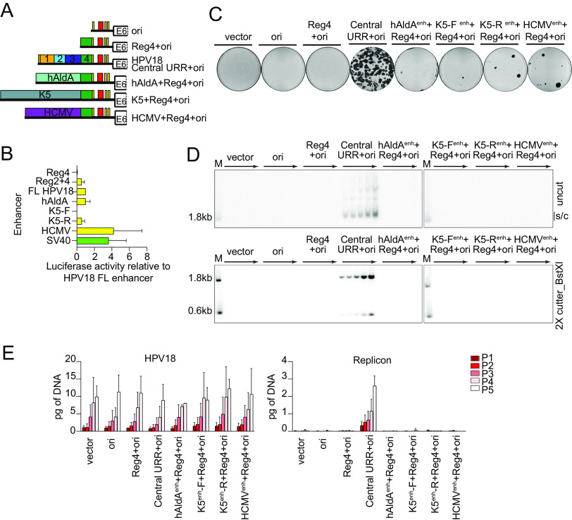
FIG 4.
Enhancer activity alone cannot complement the HPV18 central URR enhancer element for replicon maintenance. (A) Diagram of enhancer replicons. (B) Keratinocytes were transfected with luciferase reporter plasmids containing the cis elements indicated upstream from a minimal promoter, as well as a positive control (pGL4.13 expressing luciferase from the SV40 enhancer/promoter; shown in green) and a negative control (empty luciferase vector containing a minimal promoter). Cell lysates were collected 48 h after transfection, and luciferase activity was measured and normalized to the total protein concentrations for each cell lysate and the negative control plasmid. Reporter activity for each enhancer is shown relative to the HPV18 central URR enhancer. (C) Keratinocyte colonies arising from continuous G418 selection were stained with methylene blue approximately 14 days posttransfection. (D) DNA collected from five passages of cotransfected cells were analyzed by Southern blotting as described in Fig. 2C. The monomeric, supercoiled form (s/c) of the replicon is indicated. (E) HPV18 genome (left) and replicon (right) copy numbers were measured by qPCR. Error bars represent the standard deviation. The data shown are representative (panels C and D) or an average of two (panel E) biological replicates. The average of three biological replicates is shown in panel B.

