
FIG 6.
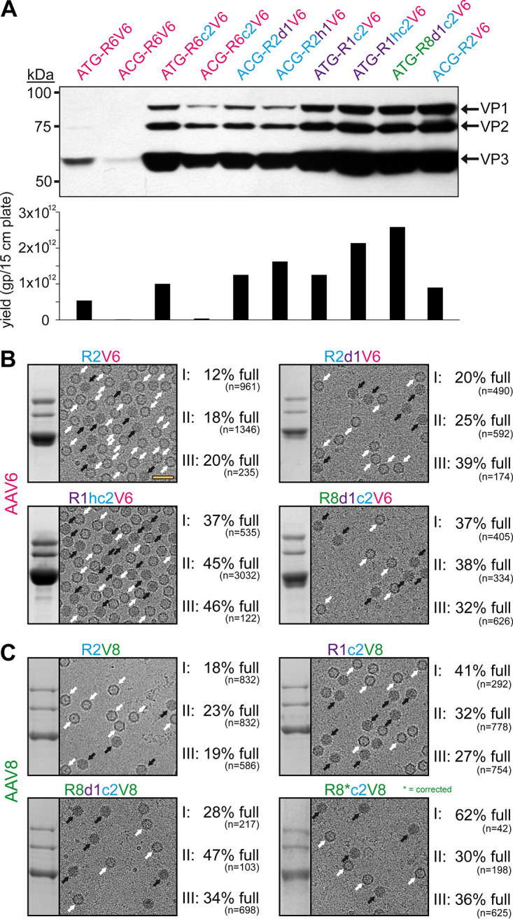
Rep hybrids enhance packaging efficiency for AAV6 and AAV8. (A) Analysis of VP expression by Western blotting and the AAV6 vector yield by qPCR after transfection of the constructs in HEK293 cells. The Western blot was probed with MAb B1. The individual VPs are indicated. (B) Analysis of the AVB-purified AAV6 vector preparations. Sections of SDS-PAGEs containing VP1, VP2, and VP3 and representative example cryo-EM micrographs are shown for each Rep variant. White arrows point to empty capsids (light appearance), and black arrows point to full capsids (dark appearance). The determined percentage of full capsids of three independently produced and purified AAV6 vector preparations are displayed with the total particle count of all micrographs collected for the individual sample. Scale bar (shown in R2V6 micrograph), 50 nm. (C) Analysis as in panel B for AAV8-Capture select affinity ligand purified AAV8 vectors.