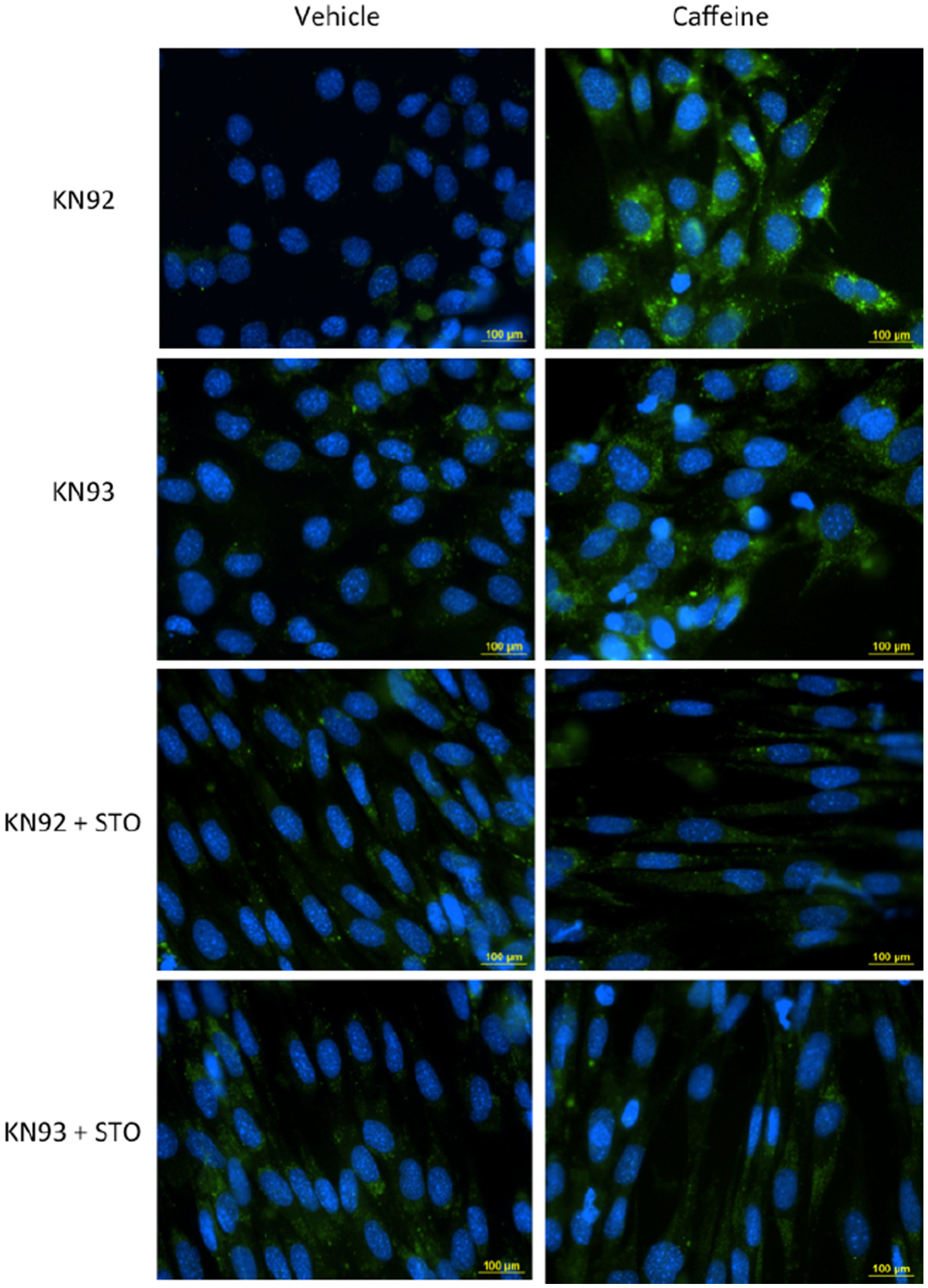
Fig. 4.

CaMKKβ is the primary mediator of AMPK-dependent autophagic vesicle accumulation in caffeine treated skeletal muscle cells. C2C12 myotubes were treated with the CaMKII inhibitor KN93 (10 μM) or its positive control KN92 (10 μM) alone or in combination with CaMKKβ inhibitor STO (30 μM) and incubated in media containing vehicle (DMSO) or 10 mM caffeine for 6 h. For the last 30 min of incubation the cells received 1 μM of Hoechst 33342 and 2 μl/ml of Cyto-ID Green (fluorescein) dye to label autophagic vesicles (autophagosomes and autolysosomes). Fluorescence microscopy analysis was conducted to examine the accumulation autophagic vesicles in each experimental treatment (n = 3). Ten individual images were taken in random locations on each coverslip (sample). The mean FITC fluorescence intensity was calculate for each sample and was used to for the quantitative assessment of autophagic vesicle accumulation between experimental treatments. Cells treated with KN92 and KN93 + 10 mM caffeine displayed a significant increase in autophagic vesicle accumulation (FITC intensity) compared to other experimental treatments (P < 0.05). The addition of STO completely inhibited the caffeine-induced increase in autophagic vesicle accumulation (P > 0.05).
