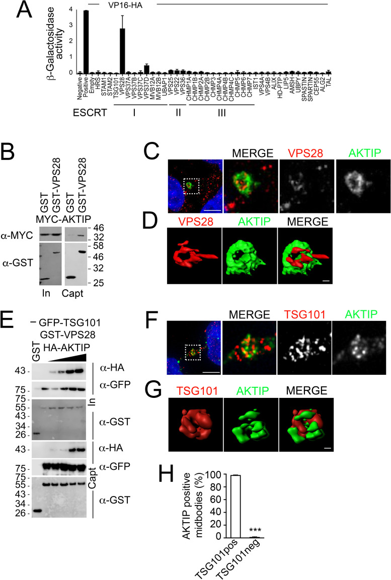
Fig 2. AKTIP is associated with the ESCRT I subunits VPS28 and TSG101.
(A) AKTIP fused to the Gal4 DNA binding domain was tested for interactions with the human components of ESCRT I, II, III, and ESCRT associated proteins fused to the VP16 activation domain by yeast two-hybrid assay. Error bars indicate the SEM from the mean of triplicate measurements. (B) Western blotting showing that AKTIP interacts with GST-VPS28 but not with GST alone. Cells were transfected with plasmids encoding the indicated fusion proteins. Purified VPS28-GST or GST alone were used to pull down interacting proteins; cell lysates and glutathione-bound fractions were then analyzed with MYC antisera. GST-pull down was repeated three times. (C) Spinning disk microscopy images of AKTIP (green) and VPS28 (red). Scale bar, 5μm. (D) 3D rendering of spinning disk imaging as in (C) showing that AKTIP and VPS28 are in proximity at midbody. VPS28 in late midbodies displays also an asymmetric protruding element. Scale bar, 0.5 μm. (E) Western blotting showing that HA-AKTIP, GFP-TSG101 and GST-VPS28 are captured together in GST pull down experiment. Cells were co-transfected with a fixed amount of plasmid encoding GFP-TSG101 (500 ng) and GST-VPS28 (1000 ng) and increasing amounts of HA-AKTIP (0, 50, 100, 500 or 1000 ng) encoding plasmid. Cell lysates and GST pull down fractions were then analyzed with GST, GFP, HA antisera. GST-pull down was repeated three times. (F) Spinning disk microscopy images of AKTIP (green) and TSG101 (red). Scale bar, 5μm. (G) 3D rendering of spinning disk imaging as in (E) showing that AKTIP and TSG101 are near each other. Scale bar, 0.5 μm. (H) Quantification of the percentage of midbodies positive both for AKTIP and TSG101 showing that the two proteins are concomitantly present at the midbody. For results in (H) at least 100 midbodies per condition were counted.

