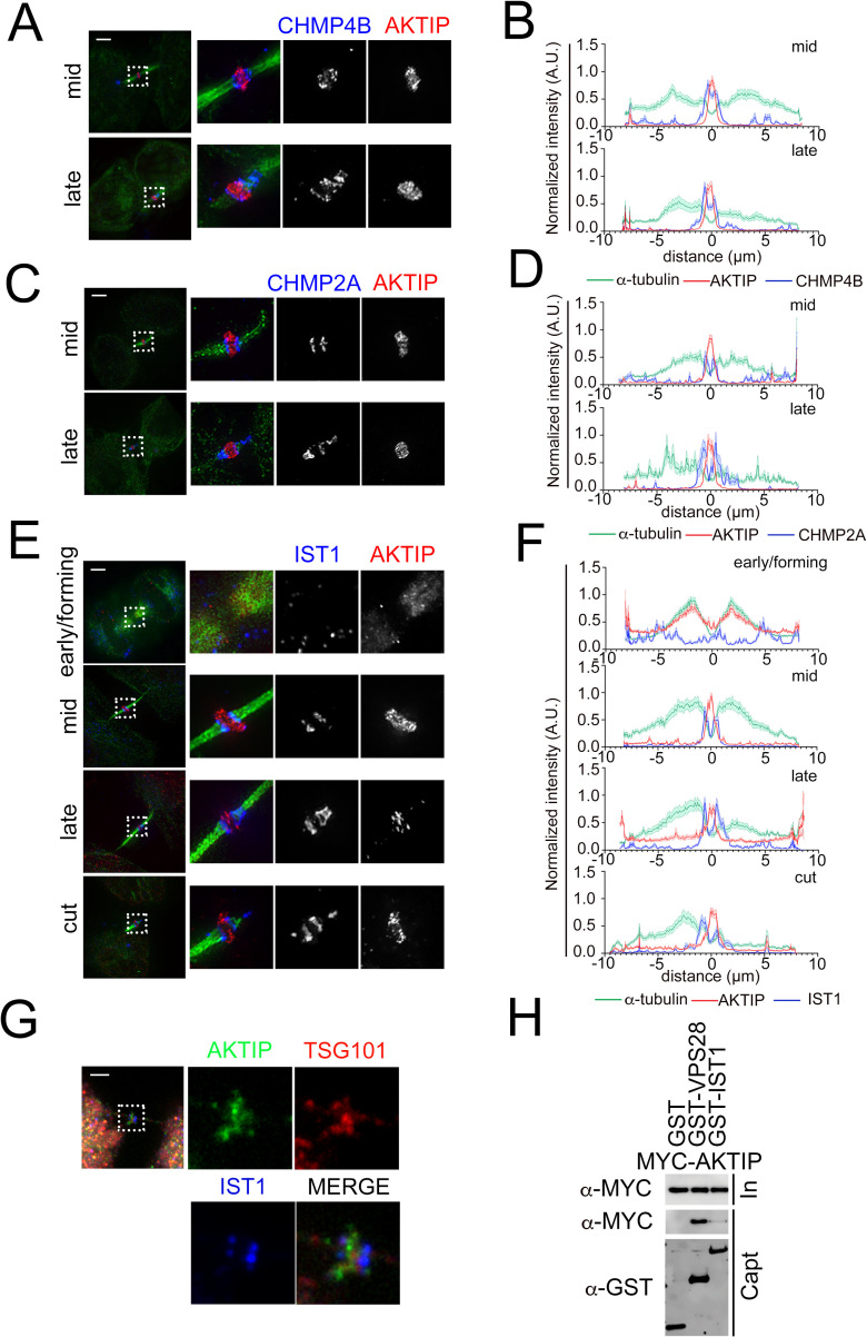
Fig 4. The supra-molecular structure of AKTIP is flanked by the rings formed by the ESCRT III components.
(A, C, E) 3D-SIM images of ESCRT III components CHMP4B (A), CHMP2A (C), IST1 (E) and AKTIP in HeLa cells. Staining with antibodies against ESCRT III (blue), AKTIP (red) and α-tubulin (green). (B, D, F) Representative fluorescence intensity profile plotted for AKTIP, ESCRT III subunits and α-tubulin along the midbodies at different stages. (B) mid, n = 6; late, n = 4. (D) mid, n = 6; late, n = 3. (F) early/forming, n = 6; mid, n = 7; late, n = 8; cut, n = 6. (G) Spinning disk microscopy images of IST1 (blue), AKTIP (green) and TSG101 (red). Scale bars, 2.5μm. (H) Western blotting showing that AKTIP interacts with GST-VPS28, but not with GST-IST1 or GST alone. Cells were transfected with plasmids encoding the indicated fusion proteins. Purified GST-VPS28 or GST-IST1 or GST alone were used to pull down interacting proteins; cell lysates and glutathione-bound fractions were then analyzed with MYC and GST antisera. GST-pull downs were repeated three times.

