FIGURE 3.

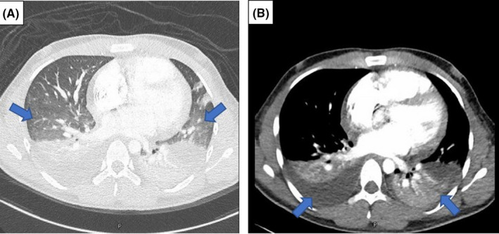
Contrast‐enhanced CT of the thorax. (A) shows bilateral ill‐defined ground‐glass opacities in the lung bases, and (A, B) depicts bilateral pleural effusion and adjacent lung collapse

Contrast‐enhanced CT of the thorax. (A) shows bilateral ill‐defined ground‐glass opacities in the lung bases, and (A, B) depicts bilateral pleural effusion and adjacent lung collapse