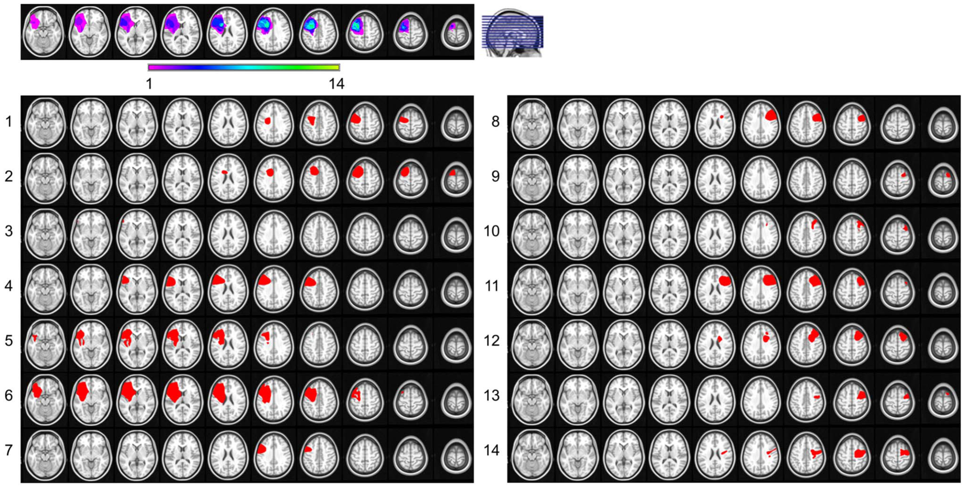
Figure 1.

pFC lesions. Reconstruction of the extent of lesion overlap for all 14 patients, normalized to the left hemisphere, shows maximal overlap in dorsolateral pFC (top). Color scale, number of patients with lesions at the specified site. pFC lesions were in the left hemisphere of seven patients and right hemisphere of seven patients (bottom). Adapted from Johnson et al. (2017).
