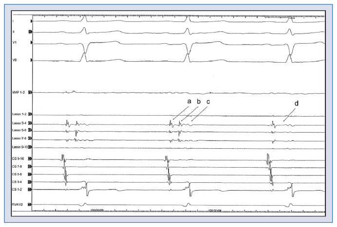
Figure 2.
Electrocardiogramm during ablation. I, II, V1, V6 = Surface-electrocardiogram, MAP = ablation catheter, Lasso 1–10 = 10 polar spiral-catheter in the left superior pulmonary vein: a — farfield atrial signal; b — pulmonary vein signal; c — farfield ventricular signal; d — no pulmonary vein signal anymore; CS 1–10 — catheter in the coronary sinus; RVA — catheter in the right ventricular apex. The 10 polar spiral-catheter is placed in the left superior pulmonary vein. During ablation around the left superior pulmonary vein, the pulmonary vein signal on the spiral-catheter disappears (b → d). This means that the vein was isolated, because there was hence, no signal passing the ablation line.

