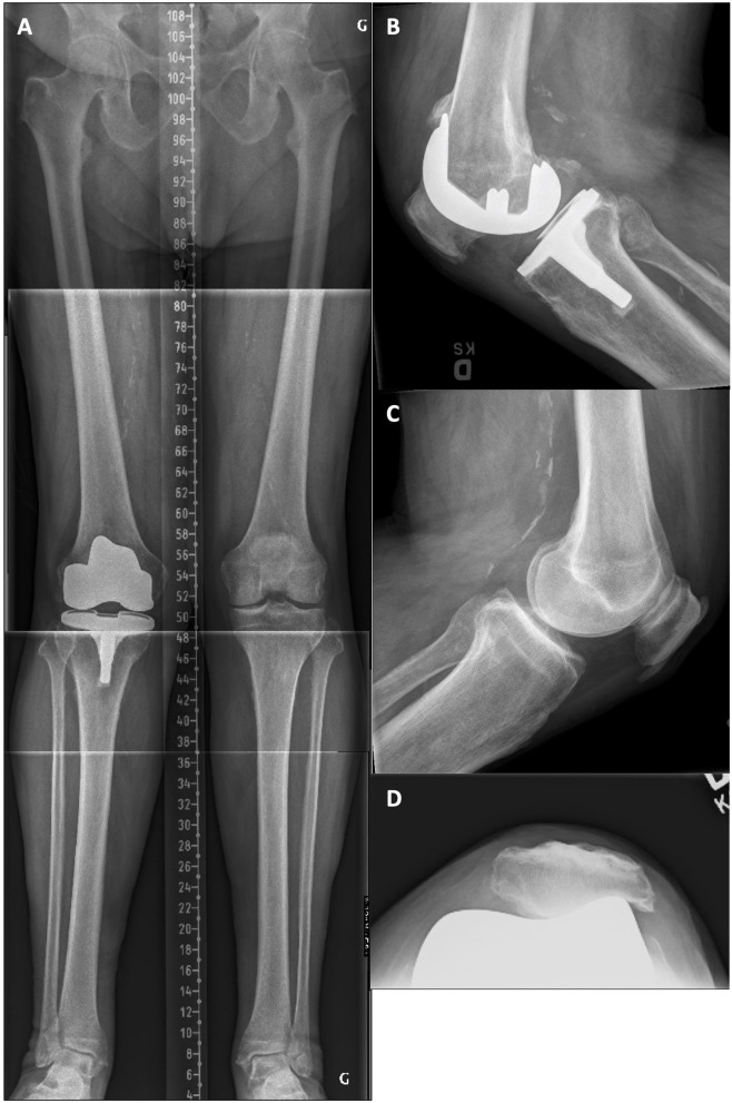
Figure 7.
(A) Pre-revision long leg standing X-Ray reveals a right implant mDFA of 4° valgus and an mPTA of 1.5° valgus (5.5° valgus aHKA). On the intact left side, the mDFA is 2.0° valgus and mPTA 0.5° varus (aHKA of 1.5° valgus). (B) Right knee lateral view where the implant tibial slope is 1.5° anterior. (C) Left knee lateral view where the native tibial slope is 8.0° posterior. (D) Right knee skyline view showing a subluxed and worn patella.

