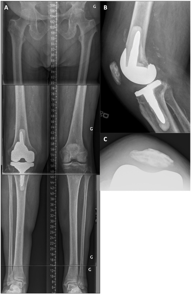
Figure 8.
(A) Post-revision standing long-leg X-ray of the right lower limb where the implant mDFA has been modified to 1.0° valgus and the mPTA at 0.5° varus (0.5° valgus aHKA). (B) Right knee lateral view where the implant tibial slope has been shifted from 1.5° anterior to 2.0° posterior. The femoral implant has also been translated posteriorly to be flush with the anterior cortex. (C) Right knee skyline view showing a well-centered resurfaced patella.

