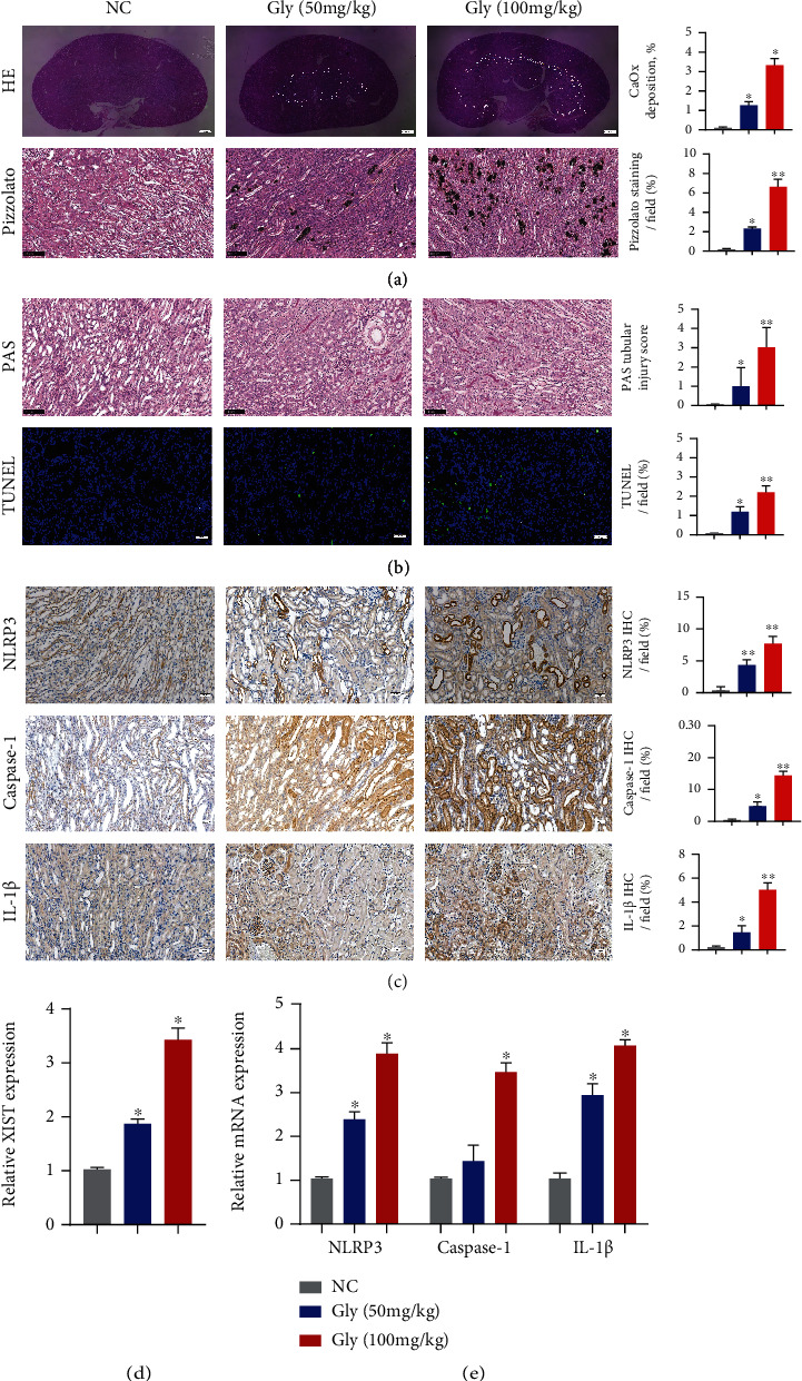
Figure 1.

XIST and NLRP3 expression was significantly increased in the CaOx nephrocalcinosis mouse model. (a) Polarizing microscopy and Pizzolato staining were performed to verify the crystal deposition after the administration of glyoxylate at varying concentrations to CaOx nephrocalcinosis model mice. (b) PAS and TUNEL staining was used to observe the kidney injury. (c) The levels of NLRP3, Caspase-1, and IL-1β and the positive ratio were detected by IHC. (d, e) The expression levels of XIST, NLRP3, Caspase-1, and IL-1β in kidney samples were determined by qPCR. The data are presented as the mean ± SD of three independent experiments. ∗P < 0.05; ∗∗P < 0.01, as assessed via Student's t-test (a–c) and one-way ANOVA (d, e).
