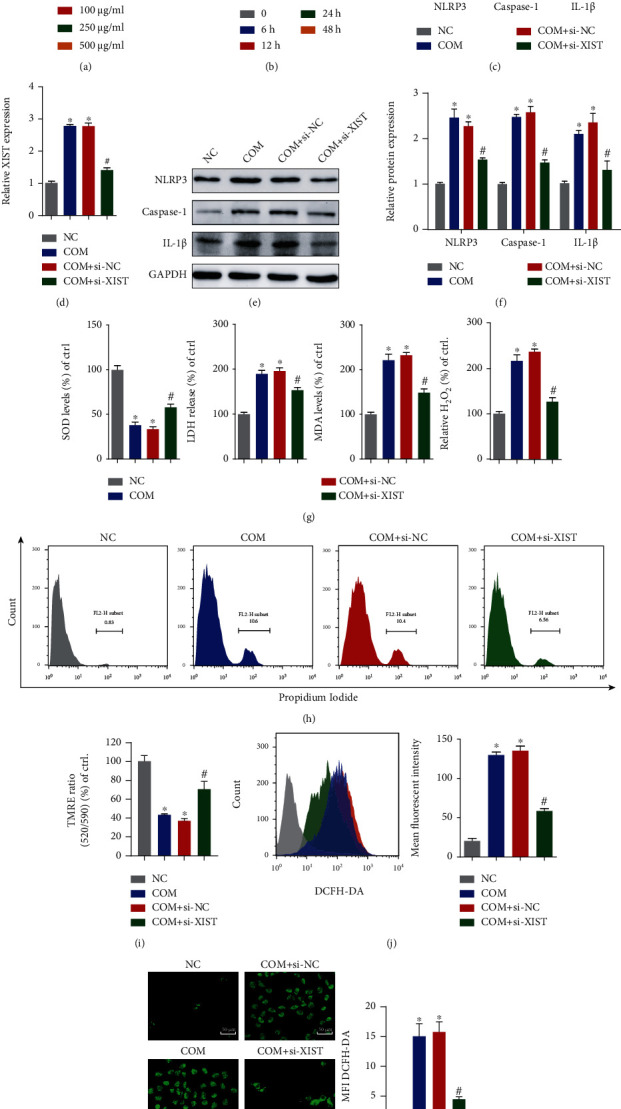
Figure 2.

Inhibition of XIST attenuated CaOx-induced renal inflammation and oxidative injury in vitro. COM at various doses (0 μg/ml, 50 μg/ml, 100 μg/ml, 250 μg/ml, and 500 μg/ml) was cocultured with HK-2 cells, and the cell viability was detected. (b) Cells were cocultured with 100 μg/ml COM for different amounts of time (0 h, 6 h, 12 h, 24 h, and 48 h), and the cell viability was examined. (c, d) qRT-PCR was used to detect the expression of XIST, NLRP3, Caspase-1, and IL-1β in HK-2 cells. (e, f) Western blotting also showed changes in the protein levels of NLRP3, Caspase-1, and IL-1β in HK-2 cells. (g) The generation of SOD, LDH, MDA, and H2O2 after different treatments was examined. (h) Flow cytometry was performed to assess cell necrosis. (i) TMRE was used to determine the mitochondrial membrane potential. Flow cytometry (j) and DCFH-DA (k) were used to detect ROS production in fluorescently labeled HK-2 cells. The data are presented as the mean ± SD of three independent experiments. ∗P < 0.05; ∗∗P < 0.01, versus the NC group, #P < 0.05, versus the COM+si-NC group, as determined by one-way ANOVA (a–d, f–k).
