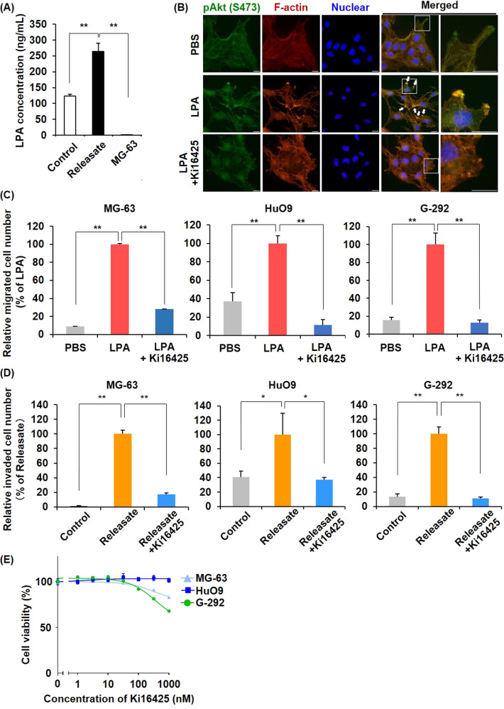
Fig. 3. LPA released from activated platelets is critical for platelet releasate-mediated osteosarcoma cell migration and invasion.
A LPA released from activated platelets was quantified by ELISA. Platelet suspensions (200 μL) and osteosarcoma cells (5 × 10⁴ cells/10 μL) were incubated for 30 min at 37 °C. The samples were collected in a 1.5-mL tube in the presence of 0.5 μM PGI2 and centrifuged at 20,000 × g for 5 min. The supernatants were then collected for cell treatments or ELISA. B Immunofluorescence staining images. MG-63 cells were starved in serum-free MEM overnight and treated with/without 10 nM LPA for 4 h. Phosphorylated Akt (green), F-actin (red) and nuclei (blue) were stained with an anti-phospho-Akt (S473) antibody, rhodamine–phalloidin reagent, and hoechst33342, respectively. Arrows indicate overlap points of phospho-Akt and F-actin. Scale bars represent 20 μm. C Effects of LPA treatment on osteosarcoma cell migration. Cells were seeded at 1 × 10⁵ cells/well in migration chambers and incubated for 4–6 h in the presence or absence of 10 nM LPA. In some cases, cells were pretreated with 100 nM Ki16425 for 1 h and seeded in migration chambers in the presence of 100 nM Ki16425. Migrated cells through the membranes were fixed and stained with crystal violet. The migrated cell number was counted and presented as percentages of the LPA values. D Effects of platelet releasate on osteosarcoma cell invasion. Cells were seeded at 1 × 10⁵ cells/well in the invasion chambers and incubated for 22–24 h in the presence or absence of platelet releasate (10 nM LPA equivalent). In some cases, cells were pretreated with 100 nM Ki16425 for 1 h and seeded in the invasion chambers in the presence of 100 nM Ki16425. Cells that had invaded through the membranes were fixed and stained with crystal violet. The invaded cell number was counted and presented as percentages of the Releasate values. E Effects of Ki16425 on cell proliferation. Cells were treated with a range of Ki16425 doses for 72 h. Cell viability was assessed using CellTiter-Glo Reagent. All data are shown as means ± SD (n = 4). **p < 0.01, *p < 0.05 as determined by Student’s t test.

