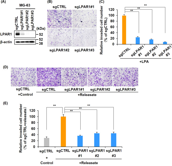
Fig. 4. LPA–LPAR1 axis is essential for platelet releasate-mediated osteosarcoma cell invasion.
A Establishment of LPAR1 knockout MG-63 cells. Cell lysates were immunoblotted with indicated antibodies. B, C Effects of LPA on MG-63/sgLPAR1#1-3 cell invasion. Cells were seeded at 1 × 10⁵ cells/well in invasion chambers and incubated for 22–24 h in the presence or absence of 10 nM LPA. Cells that had invaded through the membranes were fixed and stained with crystal violet (B scale bars represent 100 μm). The relative invaded cell number was calculated using Image J software and presented as percentages of the sgCTRL values (C). All data are shown as means ± SD (n = 4). **p < 0.01 by the Student’s t test. D, E Effects of platelet releasate on MG-63/LPAR1-KO cell invasion. Cells were seeded at 1 × 10⁵ cells/well in invasion chambers and incubated for 22–24 h in the presence or absence of platelet releasate (10 nM LPA equivalent). Invaded cells through the membranes were fixed and stained with crystal violet (D scale bars represent 100 μm). The relative invaded cell number was calculated by the Image J software and presented as percentages of the values of sgCTRL (D). All data are shown as means ± SD (n = 4). **p < 0.01 as determined by Student’s t test.

