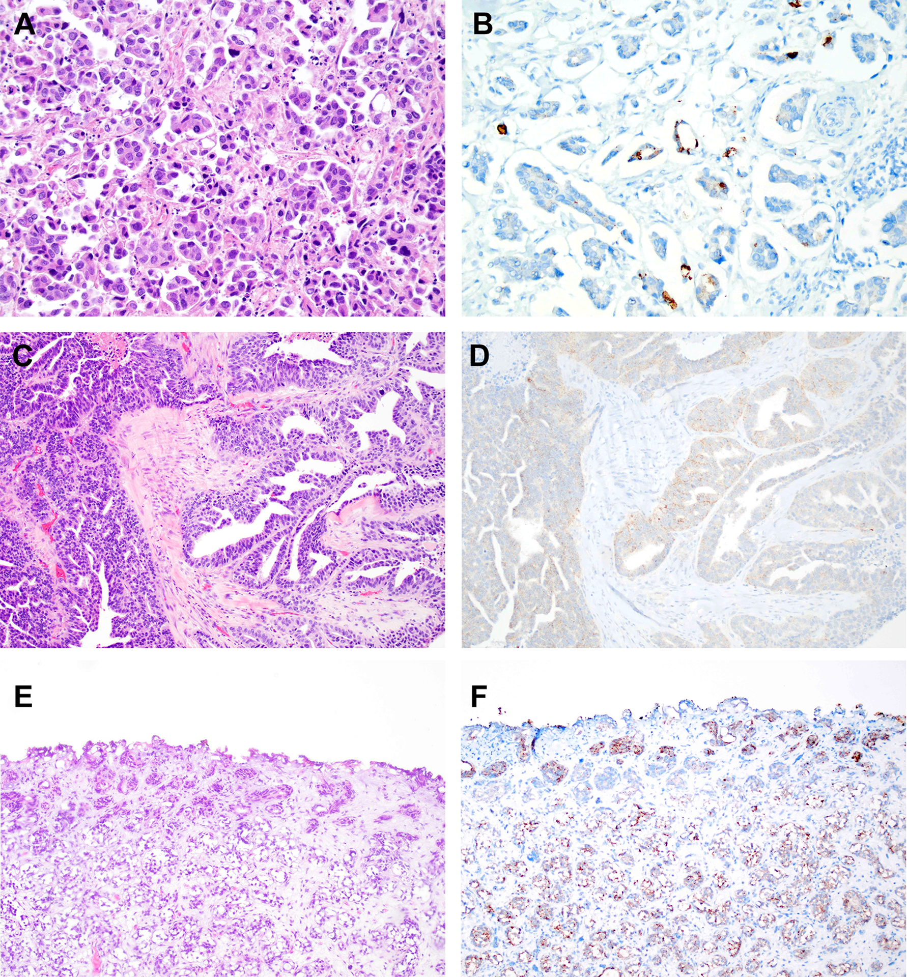
Figure 2. Immunohistochemistry for nectin-4 in micropapillary urothelial carcinoma, urothelial carcinoma with glandular differentiation, and nested urothelial carcinoma.

A. Hematoxylin and eosin (H&E) stained section of micropapillary urothelial carcinoma. B. Immunohistochemistry for nectin-4 in same tumor as in “A” showing strong membranous staining in a subset of tumor cells. C. H&E stained section of urothelial carcinoma with glandular differentiation. D. Immunohistochemistry for nectin-4 in same tumor as in “C” demonstrating weak cytoplasmic staining. E. H&E stained section of nested variant of urothelial carcinoma. F. Immunohistochemistry for nectin-4 in same tumor as in “E” demonstrating moderate staining in a subset of tumor cells.
