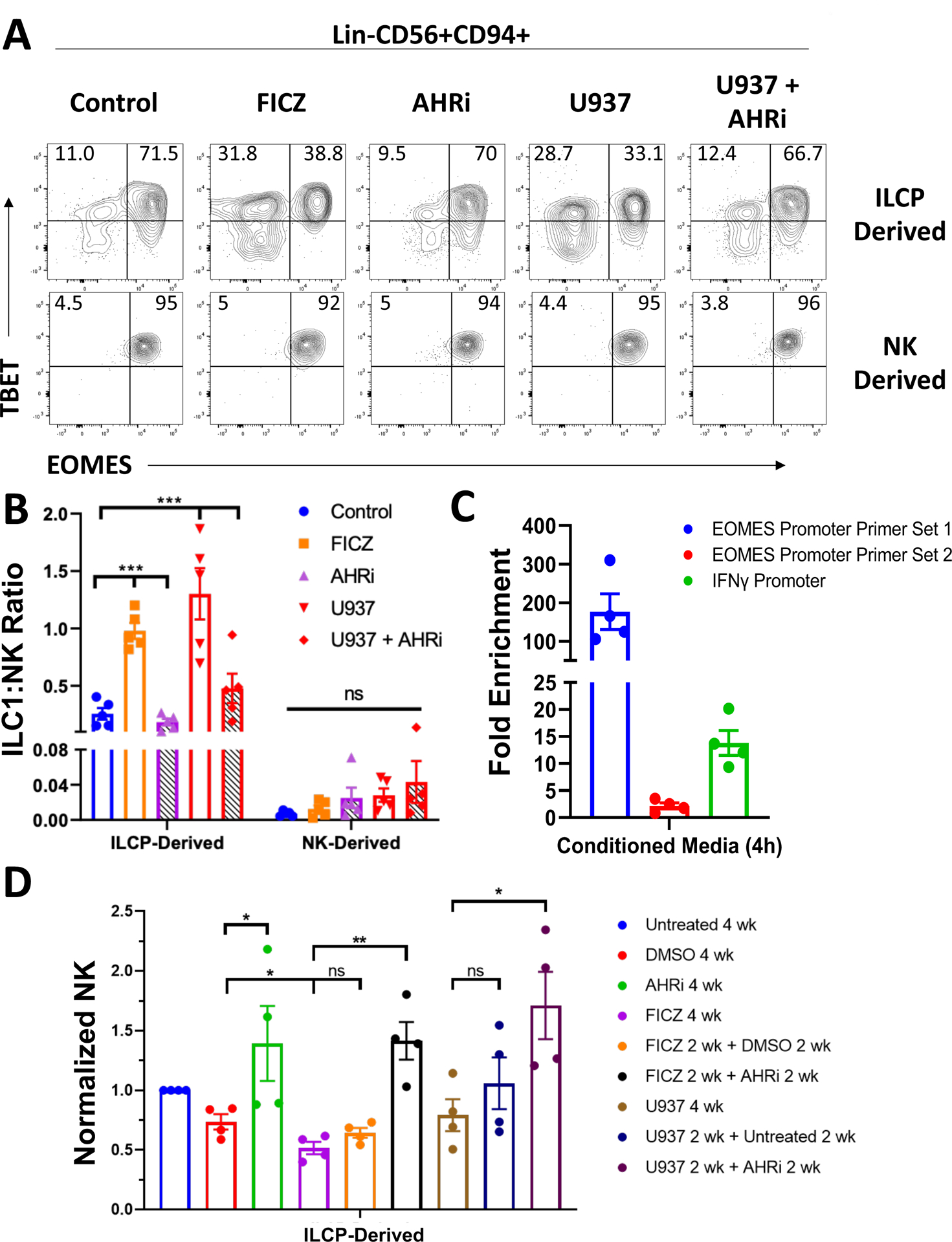
Figure 4: In vitro AHR activation promotes an ILC1 phenotype in AML.

A) Post-culture analysis of TBET and EOMES expression among Lin-CD56+CD94+ cells. ILCPs (Lin-NKp80-CD294-KLRG1-NKp44-CD94−CD16−CD117+) and CD56bright NK cells (Lin-CD56+CD94+CD16−) were isolated from normal peripheral blood of 5 independent donors and cultured with IL-7 on OP9-DL1 cells for 4 weeks. Culture conditions included: 1) treatment with the AHR agonist FICZ (30 nM), 2) the AHR inhibitor (AHRi) CH223191 (3 μM), or 3) the AML cell line U937 (± AHRi). U937 were co-cultured in transwells. Isolated populations were distributed evenly between conditions following initial cell sorting.
B) Summary data of ILC1:NK ratios across all conditions for ILCP- and NK-derived Group 1 ILCs. ANOVA was used to compare AHR agonism and AHR inhibition to control conditions for the indicated comparisons.
n=5, *p<0.05, ***p<0.001. Error bars represent ±SEM. ANOVA was used for analysis in B).
C) qPCR results of AHR ChIP pull down from peripheral blood NK cells of n=4 biological donors treated for 4 hours with AML conditioned media (from U937 cells). Fold enrichment was calculated through the equation 2−([Ct AHR ChIP]-[Ct Isotype ChIP]). Two primer sets targeting different regions of the EOMES promoter containing AHR binding motifs were tested as well as a commercially available primer set targeting the IFNG promoter (Cell Signaling Technologies).
D) Summary data of normalized NK counts for the ILCP switch culture assay. ILCPs were isolated from the peripheral blood of 4 independent donors and cultured on OP9-DL1 stromal cells with IL-7 for a total of 4 weeks. Cells were either cultured for 4 consecutive weeks in the indicated conditions or had a change in treatment after 2 weeks as indicated.
n=4, *p<0.05, **p<0.01. Error bars represent ±SEM.
